Submitted on 2024, 25th July; revised on: 2025, 16th December; accepted on: 2025, 17th December
https://doi.org/10.36253/a_h-16392
Study of the anatomy and histology of the female reproductive system of the Asian snake-eyed skink, Ablepharus pannonicus (Sauria: Scincidae)
Department of Biology, Faculty of Science, Razi University, Kermanshah, Iran
*Corresponding author. Email: r.karamiani@razi.ac.ir
Abstract. This study examines and contrasts the anatomy and histology of the female reproductive system in Ablepharus pannonicus, a member of the Scincidae family, across the spring and autumn seasons. Female specimens of A. pannonicus were collected from the northern slopes of the Shorkhah Dizah village (34°23’49.1”N 46°03’15.1”E), located 125 km west of Kermanshah Province in western Iran. After anaesthesia, the specimens were dissected in the laboratory, and their reproductive systems were processed for tissue analysis, using serial sections stained with Hematoxylin and Eosin for detailed histological examination. The findings indicate that during the spring, the ovaries of A. pannonicus contain all stages of follicular development, including primordial, mature, and ovulation-ready follicles. In contrast, the autumn samples predominantly exhibit primordial or preovulatory follicles, with a notable absence of mature follicles. These observations suggest a seasonal reproductive pattern in female A. pannonicus, characterized by significant morphological changes and heightened activity within the ovaries and genital tracts during spring. As the ambient temperature drops and the reproductive season concludes, gonadal size and activity decrease, leading to the absence of reproductive activity in autumn. It is hypothesized that during this period, the animals engage in feeding and fat storage in preparation for hibernation.
Keywords. Reproductive cycle, Ablepharus pannonicus, reproductive system, ovary.
Contents
The exploration of reproductive diversity among lizards offers a rich tapestry of biological phenomena for scientific inquiry. These reptiles display a remarkable array of reproductive strategies: from oviparity to viviparity, prolific to sparse offspring production, and varied levels of parental investment. Some species are known to lay eggs immediately post-shell formation, while others retain shelled eggs for prolonged periods before laying. Remarkably, viviparity has independently evolved numerous times within squamates, and certain species even exhibit parthenogenesis. These reproductive variations offer a glimpse into the captivating complexity of lizard reproductive ecology (Rheubert et al., 2014).
Lizards typically exhibit one of three reproductive cycles: constant, associated, or dissociated. The constant cycle is characterized by nearly year-long gonadal activity. In contrast, the associated and dissociated cycles feature a discontinuous mating season. The associated cycle is marked by a surge in gonadal activity just before the mating season, synchronously in both sexes, obviating the need for sperm storage due to its ready availability (Censky, 1995; Huang, 1997). Conversely, the dissociated cycle is defined by diminished gonadal activity during the mating season, with a peak in the non-mating period. Notably, male gonadal activity is briefer compared to females, necessitating the storage of sperm within the female reproductive tract for subsequent fertilization (Torki, 2006).
The oviductal structure in lizards exhibits a conserved morphology, characterized by three distinct regions identifiable across species. These regions, extending from the caudal to the cranial end, comprise the nonglandular uterus – commonly referred to as the vagina – the glandular uterus, typically known as the uterus, and the infundibulum (Siegel et al., 2011). Female lizards possess bilateral oval ovaries anchored to the dorsal body wall via a slender mesovarium. The ovarian cortex is surrounded by the tunica albuginea, and is lined by a simple squamous epithelium (Klosterman, 1983; Guraya, 1989). Oogenesis occurs within the germinal beds situated dorsally on the ovary, proximal to the mesovarium. These beds harbor proliferating oogonia, undifferentiated somatic cells, oocytes encased by a few somatic cells, and primary follicles (Jones and Guillette, 1982; Klosterman, 1983). The ovarian follicles, serving as the functional units of the ovary, are organized into a hierarchical structure, including primordial, primary, secondary, previtellogenic, vitellogenic, and preovulatory follicles. This follicular hierarchy is a well-defined feature of reptilian ovarian architecture (Guraya, 1989; Etches and Petitte, 1990).
The skink Ablepharus pannonicus (see Fig. S1) inhabits a range of environments including semi-desert and desert areas, steppe habitats, and mountainous terrains. Its geographical distribution extends from the Middle East to Central and South Asia, covering countries such as Iraq, Iran, Jordan, Syria, Afghanistan, Pakistan, India, Kyrgyzstan, and Uzbekistan (Karamiani, 2018). Despite the breadth of studies on various aspects of this species, there is a notable gap in the literature regarding the anatomical and histological characterization of the female reproductive system. Addressing this lack of information, the present study provides the first detailed description of the anatomy and histology of the female reproductive system of A. pannonicus, offering insights into a previously unexplored aspect of this widely distributed lizard.
This research was conducted in the natural surroundings of the village of Shorkhah Dizah (34°23’49.1”N 46°03’15.1”E), located 125 km west of the city of Kermanshah, in Kermanshah Province, western Iran. Sampling was conducted in the spring (April–May) and autumn (October–November) of 2021, with four adult female specimens of A. pannonicus collected in each season. These two sampling periods were selected to capture contrasting reproductive stages: spring representing peak ovarian activity and autumn reflecting a regressed state, consistent with seasonal reproductive patterns reported in scincid lizards (Sever and Hopkins, 2004; Vergilov et al., 2018). According to long-term climate normals (1981–2010) reported by the Iran Meteorological Organization (IRIMO), average temperatures in the study area range from 15-22 °C in spring and 9-17 °C in autumn. Rainfall is moderate in spring but substantially lower in autumn (Iran Meteorological Organization, 2021).
Upon arrival at the laboratory, the specimens underwent anaesthesia using diethyl ether-soaked cotton. Subsequently, we euthanised them through intracoelomic injection of sodium pentobarbital, following the animal care guidelines approved by Southeastern Louisiana University Animal Care and Use Committee (Rheubert et al., 2020). Next, we meticulously recorded morphometric parameters, including snout–vent length (SVL) and body mass. A longitudinal incision was made on the ventral surface of the specimens, allowing for the removal of the digestive tract. This surgical approach exposed the reproductive system, which we then photographed for subsequent analysis. The entire oviductal tract was carefully dissected and weighed using a digital analytical balance. Using this data, we calculated the gonadosomatic index (GSI), providing a quantitative measure of reproductive investment (Jacobson, 2007). The GSI was calculated as GSI = (ovary weight / body weight) × 100, reflecting the proportional reproductive investment of each individual. After the oviductal tract was removed from the lizard’s body, the samples were immediately placed in 10% formalin for 72 h. Subsequently, the samples were dehydrated in a series of ethanol solutions: 60% for 60 min, 70% for 30 min, 80% for 30 min, and 96% for 120 min. For clearing, the samples were placed in three containers containing xylene, each for 30 min. The samples were then embedded in paraffin in an incubator set at 58°C. In this step, three containers of paraffin were used, and the capsules containing the tissue samples were placed in each container for 120 min to allow the paraffin to replace the xylene in the tissue. L-shaped metal molds, known as Leuckhard molds, were used for molding. The mold was adjusted to the appropriate size, and molten paraffin was then poured into the mold. The samples were subsequently placed vertically inside it. In the next step, the blocks were separated from the mold, resulting in a molded block for each individual. A rotary microtome (CUT SLEE 4060) was used to obtain transversal and longitudinal sections from paraffin blocks containing the entire oviductal tract. Sections were prepared from multiple regions along the tract, and representative slides were selected for histological analysis. The sections were stained using hematoxylin-eosin staining (Suvarna et al., 2018).
The female reproductive system in Ablepharus pannonicus comprises a pair of ovaries and an oviduct. The oviducts traverse past the kidneys and terminate in the cloaca (Fig. 1A, B). The ovaries are situated within the abdominal cavity as whitish glandular structures, attached to the dorsal wall by the mesovarium. The ovarian surface exhibits small and large protrusions due to the presence of follicles at various stages of maturity. Each ovary is enveloped by a thin covering known as the tunica albuginea. During the spring, all types of follicles (especially vitellogenic and preovulatory follicles) are observable in the ovarian sections (Fig. 1C). However, in autumn samples, mature follicles are scarce, and the predominant follicles are in the primary, secondary, or previtellogenic stage (Fig. 1D).
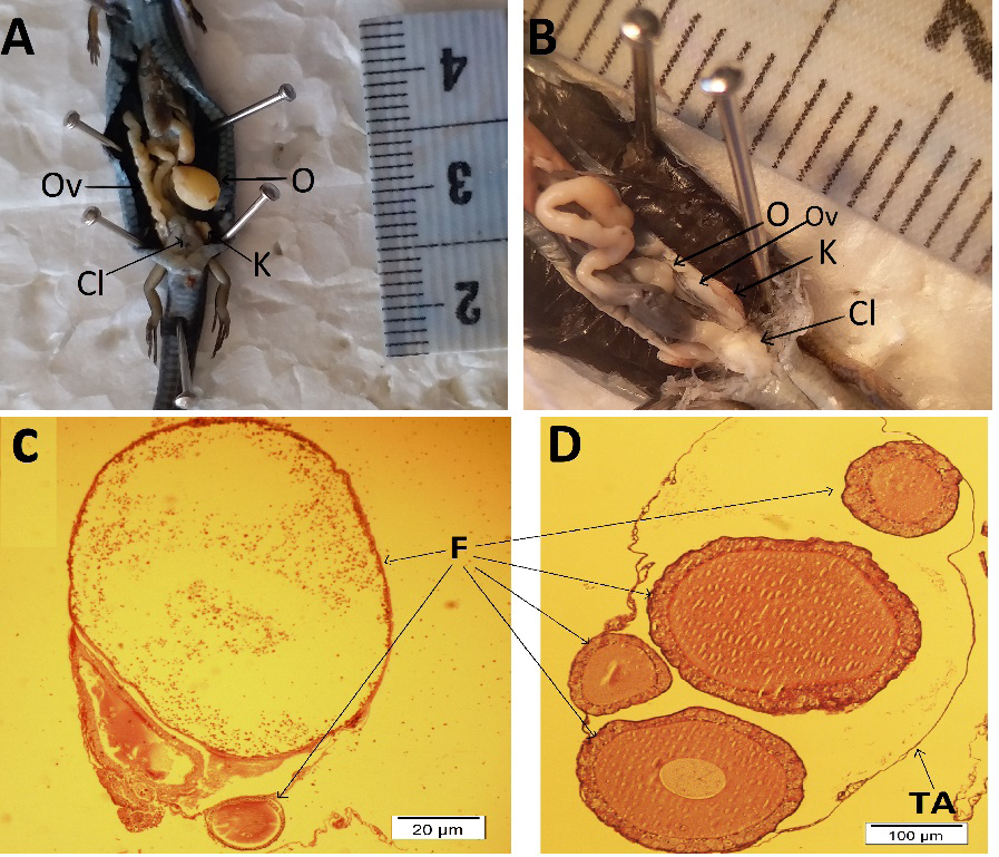
Microscopic examination of the previtellogenic follicle wall reveals at least two distinct cell types – large and small cells (see Fig. S2). The ovarian and body weight data, and gonadosomatic index data collected from each season are presented in Table S1. No notable structural differences were observed in the oviductal anatomy or histology between spring and autumn specimens. Moving along the oviduct, the most anterior portion is referred to as the infundibulum. The epithelial cells lining its proximal region exhibit a squamous to cuboidal morphology. Toward the distal region, the cellular height increases, culminating in a cylindrical form. Notably, certain cells in the distal infundibulum display abundant cilia (Fig. 2A, B).
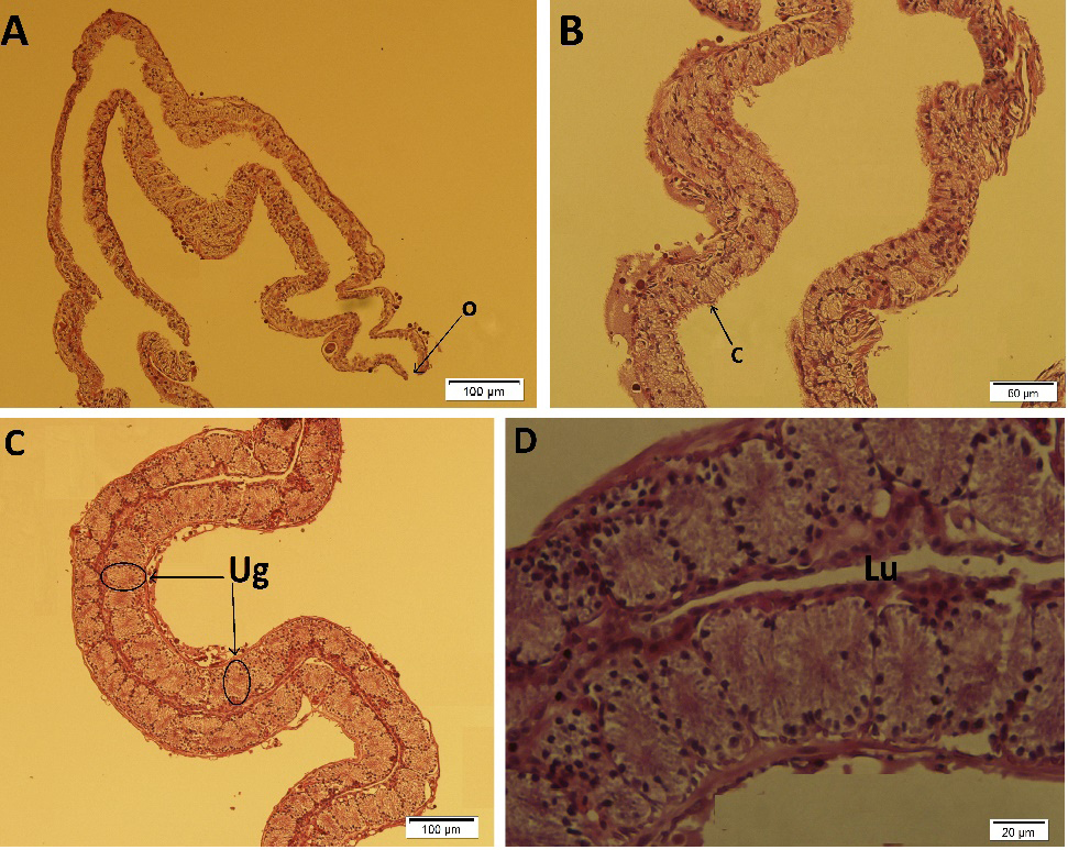
The glandular uterus constitutes the central segment of the oviduct in A. pannonicus. Its wall is densely populated with mucous glands, which are acinar in configuration and embedded within the tissue. The predominantly columnar cells of these glands play a crucial role in eggshell formation through their secretions (Fig. 2C, D).
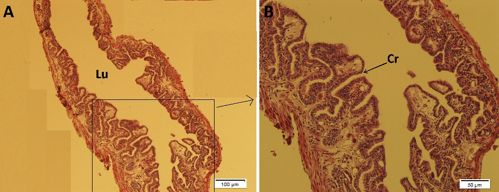
The most posterior region of the oviduct is the non-glandular uterus, connecting the glandular uterus to the urodeum of the cloaca. In this area, numerous crypts are present, their abundance increasing as one approaches the cloacal urodeum (see Fig. 3 and Fig.S3).
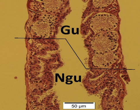
Unlike the glandular uterus, this region lacks secretory glands, allowing for clear demarcation (see Fig. 4). The number of Germinal Beds (GBs) per ovary exhibits both intra- and inter-specific variation, ranging from one to six among lizard species (Jones et al., 1979; Radder et al., 2008). Studies have posited a correlation between the number of GBs per ovary, clutch size, and breeding frequency. Jones and Guillette (1982) identified a pattern linking the number of GBs to clutch size. Typically, species that undergo monoallochronic ovulation – ovulating a single egg alternately from each ovary, as seen in the Dactyloidae family (Jones et al., 1979) – and those with low fixed clutch sizes and monoautochronic ovulation – releasing a single egg simultaneously from both ovaries, such as in Gekkota (Jones and Summers, 1984) and Gymnophtalmidae (Vitt, 1982) – possess one GB per ovary. Conversely, species with high fecundity, characterized by polyautochronic ovulation and multiple large clutches, exhibit two or more spatially distinct GBs in each ovary (Radder et al., 2008). In all A. pannonicus specimens examined during the spring, GBs were indistinct. However, the presence of a single follicle poised for ovulation in both ovaries suggests a monoallochronic ovulation pattern.
Within the walls of previtellogenic follicles in certain lizards, granulosa cells of varying types are discernible. For instance, in Ablepharus kitaibelii’s previtellogenic follicles, three granulosa cell types – small, large, and pyriform – are identifiable (Vergilov et al., 2018). In this study, two cell types – large and small – were observed in A. pannonicus’s previtellogenic follicle walls, yet pyriform cells were absent.
In species like Eumeces egregius (Schaefer and Roeding, 1973) and Scincella lateralis (Sever and Hopkins, 2004), the non-glandular uterus contains numerous ciliated crypts functioning as sperm storage sites. Examination of the non-glandular uterus of A. pannonicus revealed numerous crypts, similar to those reported in the species mentioned above. However, no sperm was detected in specimens collected during either spring or autumn. This absence may reflect phylogenetic differences among scincid lizards or limitations of our sample size, rather than conclusively indicating that the crypts are not used for sperm storage.
Glandular uterine glands play a pivotal role in egg membrane formation via their secretions. These glands exhibit considerable morphological diversity across lizard species. In Plestiodon obsoletus, the glands are tubular (Guillette Jr. et al., 1989), while in Chalcides chalcides, they are tubular with simple cuboidal epithelium (Blackburn et al., 1998), and in Chalcides ocellatus, they are simple alveolar (Corso et al., 2000). In these species, secretions are expelled through an end duct to the uterus’s glandular lining. The glands in the glandular uterine wall of A. pannonicus were acinar with columnar epithelial cells in specimens from both spring and autumn. While no seasonal structural differences were noted, potential variations in functional activity cannot be ruled out.
The infundibulum, featuring numerous crypts in lizards like Acanthodactylus scutellatus and Hemidactylus turcicus, is recognized as a sperm storage site (Bou-Resli et al., 1981; Eckstut et al., 2009). However, comprehensive documentation of sperm storage in this region among skinks is lacking. In A. pannonicus, as in other skinks, sperm was not detected in the infundibulum.
These findings suggest that reproduction in female A. pannonicus is seasonal, as indicated by significant morphological changes in the gonads and increased activity in the genital ducts during spring. This conclusion is further supported by a marked increase in the gonadosomatic index (GSI) observed in spring-collected specimens compared to those collected in autumn, reflecting a higher reproductive investment during the active breeding season. As the ambient temperature declines, a noticeable seasonal reduction in both gonadal size and GSI is evident, corresponding with the cessation of reproductive activity. Consequently, this lizard exhibits no signs of sexual activity during the autumn season. This seasonal variation in gonadal condition and reproductive readiness aligns with regional climatic patterns, where average daily temperatures range from 15–22 °C in spring and decrease to 9–17 °C in autumn in the mountainous areas south of Kermanshah Province.
Acknowledgments
We wish to thank Mr. Morad Besharati for assisting us with field work. This study was conducted in accordance with the ethical standards of Razi University (Ethics Approval Code: IR.RAZI.REC.1399.003).
Supplementary material
Supplementary material associated with this article can be found at https://oaj.fupress.net/index.php/ah/article/view/16392/15293.
References
Blackburn, D.G., Kleis‐San Francisco, S., Callard, I.P. (1998): Histology of abortive egg sites in the uterus of a viviparous, placentotrophic lizard, the skink Chalcides chalcides. J. Morphol. 235: 97-108.
Bou-Resli, M.N., Bishaw, L.F., Al-Zaid, N.S. (1981): Observations on the fine structure of the sperm storage crypts in the lizard Acanthodactylus scutellatus hardyi. Arch. Biol. (Bruxelles) 92: 287–298.
Censky, E.J. (1995): Reproduction in two lesser antillean populations of Ameiva plei (Teiidae). J. Herpetol. 29: 553-560.
Corso, G., Delitala, G.M., Carcupino, M. (2000). Uterine morphology during the annual cycle in Chalcides ocellatus tiligugu (Gmelin) (Squamata: Scincidae). J. Morphol. 243: 153-165.
Eckstut, M.E., Lemons, E.R., Sever, D.M. (2009): Annual dynamics of sperm production and storage in the Mediterranean gecko, Hemidactylus turcicus, in the southeastern United States. Amphib.-Reptil. 30: 45-56.
Etches, R.J., Petitte, J.N. (1990): Reptilian and avian follicular hierarchies: Models for the study of ovarian development. J. Exp. Zool. 256: 112-122.
Guillette Jr, L.J., Fox, S.L., Palmer, B.D. (1989): Oviductal morphology and egg shelling in the oviparous lizards Crotaphytus collaris and Eumeces obsoletus. J. Morphol. 201: 145-159.
Guraya, S.S. (1989): Ovarian follicles in reptiles and birds. Zoophysiology 24: XIII-285.
Huang, W.S. (1997): Reproductive cycle of the oviparious lizard Japalura brevipes (Agamidae: Reptilia) in Taiwan, Republic of China. J. Herpetol. 31: 22-29.
Iran Meteorological Organization. 2021. Climate Normals (1981-2010). Iran Meteorological Organization, Tehran, Iran.
Jacobson, E.R. (2007): Biology, medicine, and surgery of reptiles. Boca Raton, Florida, CRC Press.
Jones, R.E., Fitzgerald, K.T., Duvall, D., Banker, D. (1979): On the mechanisms of alternating and simultaneous ovulation in lizards. Herpetologica 35: 132-139.
Jones, R.E., Guillette Jr, L.J. (1982): Hormonal control of oviposition and parturition in lizards. Herpetologica 38: 80-93.
Jones, R.E., Summers, C.H. (1984): Compensatory follicular hypertrophy during the ovarian cycle of the house gecko, Hemidactylus frenatus. Anat. Rec. 209: 59-65.
Karamiani, R., Rastegar-Pouyani, N., Rastegar-Pouyani, E. (2018): Modeling the past and current distribution and habitat suitability for Ablepharus grayanus and A. pannonicus (Sauria: Scincidae). Asian Herpetol. Res. 9: 56-64A.
Klosterman, L.L. (1983): The ultrastructure of germinal beds in the ovary of Gerrhonotus coeruleus (Reptilia: Anguidae). J. Morphol. 178: 247-265.
Radder, R.S., Pizzatto, L., Shine, R. (2008): Morphological correlates of life-history variation: Is lizard clutch size related to the number of germinal beds in the ovary? Biol. J. Linn. Soc. 94: 81-88.
Rheubert, J.L., Siegel, D.S., Trauth, S.E. (2014): Reproductive biology and phylogeny of lizards and tuatara. Boca Raton, Florida, CRC Press.
Rheubert, J., Pasternak, M.A., Ely, M., Siegel, D.S., Trauth, S.E., Gribbins, K.M., Sever, D.M. (2020): Seasonal histology and ultrastructure of the urogenital system in two sympatric lizards. J. Zool. 310: 273-286.
Schaefer, G.C., Roeding, C.E. (1973): Evidence for vaginal sperm storage in the mole skink, Eumeces egregius. Copeia 1973: 346-347.
Sever, D.M., Hopkins, W.A. (2004): Oviductal sperm storage in the ground skink Scincella laterale Holbrook (Reptilia: Scincidae). J. Exp. Zool. A Comp. Exp. Biol. 301: 599-611.
Siegel, D.S., Miralles, A., Chabarria, R.E., Aldridge, R.D. (2011): Female reproductive anatomy: Cloaca, oviduct, and sperm storage. In: Reproductive biology and phylogeny of snakes, pp. 347-409. Aldridge, R. D., Sever, D. M., Eds, Florida, CNC Press
Suvarna, K.S., Layton, C., Bancroft, J.D. (2018): Bancroft’s theory and practice of histological techniques. Elsevier Health Sciences.
Torki, F. (2006): Spermatogenesis in the agama Trapelus lessonae (Agamidae: Reptilia) in the central Zagros Mountains, Iran. Zool. Middle East 38: 21-28.
Vergilov, V.S., Necheva, V.G., Zlatkov, B.P. (2018): Reproduction of snake-eyed skink Ablepharus kitaibelii (Bibron & Bory de Saint-Vincent, 1833) (Squamata: Scincidae) in Bulgaria. Acta Zool. Bulg. 70: 507-516.
Vitt, L.J. (1982): Sexual dimorphism and reproduction in the microteiid lizard, Gymnophthalmus multiscutatus. J. Herpetol. 16: 325-329.