https://doi.org/10.36253/a_h-18382
Submitted on: 2025, 3th August; revised on: 2026, 19th January; accepted on: 2026, 22th January.
Detection of CMTV-like ranavirus following a Rana temporaria mass mortality event in a northern Italian alpine lake
Istituto Zooprofilattico Sperimentale del Piemonte, Liguria e Valle d’Aosta, Via Bologna 148, 10154 Torino, Italy
# These authors share first authorship
§ These authors share last authorship
*Corresponding author. Email: matteoriccardo.dinicola@izsplv.it
Abstract. High-mountain lakes are vulnerable to climatic and anthropogenic stressors, and infectious disease may further exacerbate impacts on alpine communities, particularly during seasonal temperature peaks. In August 2024, a mass mortality event of common frogs (Rana temporaria) occurred in a high-altitude lake in the Cottian Alps (Piedmont, Italy). During a one-hour survey, 78 dead frogs were recorded; nine carcasses (all adult males) were sampled for diagnostic tests. Six showed ventral red discolouration and three of them had ulcerative lesions of the digits. Eight out of nine vitreous humour samples were culture-positive, with isolates including Hafnia alvei, Acinetobacter guillouiae, Acinetobacter proteolyticus, and Serratia proteamaculans. PCR screening of skin and pooled organs detected ranavirus in four out of nine frogs, while Batrachochytrium dendrobatidis and herpesvirus tested negative. Phylogenetic analysis of sequenced major capsid protein and DNA polymerase fragments grouped the virus within the CMTV-like clade, with high similarity to reference sequences. This represents the first geolocated detection of a CMTV-like ranavirus in free-ranging amphibians in Italy. Although the advanced state of decomposition precluded histopathological evaluation and causality cannot be conclusively established, the concordance between molecular detection and gross lesions consistent with ranaviral infection supports a plausible role of ranavirus in the observed die-off. Our findings highlight the need for targeted surveillance in Italy’s alpine amphibians, including environmental DNA sampling and screening of non-native fish. Given ecological simplification and short reproductive seasons at high altitude, longitudinal monitoring is advisable to assess persistence, seasonality and potential spillover across life stages and sympatric species.
Keywords. Acinetobacter, CMTV, Cottian Alps, disease ecology, emerging infectious diseases, Germanasca Valley, outbreak, Ranavirus.
Contents
Field inspection and gross pathology
Sequencing and phylogenetic analysis
Field observations and gross clinical findings
Laboratory findings and phylogenetic analysis
Introduction
High‐mountain lakes are typically small, oligotrophic waterbodies located above the tree line, characterised by extended winters, brief ice‐free periods, and extremely low nutrient levels. Their clear, cold waters support simplified biological communities, and the biodiversity of these ecosystems usually decreases with altitude, with communities composed of specialised and highly adapted taxa (Tiberti et al., 2014a; Pastorino and Prearo, 2020; Pastorino et al., 2024).
Despite their apparent isolation, alpine lakes across Europe face multiple anthropogenic stressors that can amplify ecosystem vulnerability: climate warming, especially at higher elevations, leads to increased water temperatures, reduced ice‐cover duration, altered seasonal mixing regimes, and modified hydrological cycles (Råman Vinnå et al., 2021); pastoralism can drive nutrient enrichment and increased organic loading, particularly under high grazing pressure (Tiberti et al., 2014b); atmospheric transport introduces persistent organic pollutants and contaminants of emerging concern, resulting in chronic exposure risks to native biota (Machate et al., 2023; Pastorino et al., 2024); the intentional introduction of alien fish species, salmonids in particular, significantly alters indigenous amphibian and macroinvertebrate communities via direct predation (Tiberti and von Hardenberg, 2012), and may also pose risks by facilitating the spread of pathogens, including ranaviruses (Price et al., 2017); recreational tourism and water abstraction activities add pressure through habitat disturbance, direct contamination, and hydrological alterations (Pastorino and Prearo, 2020).
Although infectious diseases were long overlooked among the primary drivers of biodiversity loss, growing evidence indicates their significant role in wildlife population declines (Smith et al., 2006, 2009). Indeed, pathogens can trigger rapid population reductions or local extinctions, particularly when acting in synergy with climate change and other anthropogenic pressures (Fisher et al., 2012; Hoberg and Brooks, 2015; Di Nicola et al., 2025). Amphibians are exemplary in this context, having experienced some of the most dramatic disease-driven declines among vertebrates, notably linked to emerging pathogens such as the chytrid fungi (Batrachochytrium dendrobatidis and B. salamandrivorans) and ranaviruses, which have caused mass mortalities worldwide (e.g., Price et al., 2014, 2017; Fisher and Garner, 2020; Hartmann et al., 2022; Akçakaya et al., 2023; Luedtke et al., 2023; Schilliger et al., 2023). Such dynamics could be particularly relevant in alpine lake ecosystems, where amphibians often represent the dominant native vertebrate fauna, a status they would frequently hold exclusively if not for the presence of introduced fish (Catalan et al., 2017). In high-altitude habitats, the combination of environmental harshness, reduced ecosystem complexity, and short reproductive seasons may increase host vulnerability and limit population resilience following disease outbreaks. Moreover, climatic and anthropogenic pressures can interact with pathogens, potentially amplifying disease dynamics and impacts in amphibian populations at higher altitudes (Bosch et al., 2007; Knapp et al., 2011).
One of the most frequent amphibians in alpine lake ecosystems is the common frog, Rana temporaria Linnaeus, 1758, which is among the most widespread Palearctic amphibians, ranging from the Iberian Peninsula to western Siberia. In southern Europe, its distribution is largely confined to montane and submontane habitats, where it breeds in wetlands at elevations of up to 2800 metres in the Alps and Pyrenees (Tiberti and von Hardenberg, 2012; Di Nicola et al., 2021; Ilić et al., 2024). At higher altitudes, R. temporaria life cycle is shaped by short summers, requiring larval development to occur within a limited time window (Bison et al., 2021), which may increase its vulnerability to phenological disruptions and to population turnover caused by disease outbreaks.
Notably, ranaviruses (large double-stranded DNA viruses of the Iridoviridae family infecting ectothermic vertebrates; Jancovich et al., 2015), have been directly implicated in mass mortality events (MMEs) within R. temporaria populations inhabiting high-altitude lakes: Miaud et al. (2016) reported three outbreaks in the Mercantour National Park (French Alps), where molecular diagnostics confirmed the presence of a ranavirus (common midwife toad virus, CMTV) in all dead frogs, with near-complete die-offs of both larvae and adults. Similar MMEs involving montane amphibians were also documented in Spain, in the Cantabrian Mountains, where R. temporaria was recorded at several affected sites and CMTV-like ranavirus was detected in at least one individual (Price et al., 2014). In the Mercantour area, CMTV-like DNA was also detected throughout an activity season in live R. temporaria tadpoles, with infection peaking during a MME and persisting in survivors until metamorphosis; adults, in contrast, showed only transient infection and no substantial die-off was observed (Miaud et al., 2019). Long-term monitoring in the Cantabrian Mountains further indicated persistent negative effects of ranavirus outbreaks, although impacts on common frogs specifically appeared less severe compared to other sympatric amphibians (Bosch et al., 2021). In the French Pyrenees, a Frog Virus 3 (FV3)-like ranavirus was sporadically detected in dead amphibians, including R. temporaria, collected during chytridiomycosis-driven die-offs, but its contribution to mortality remains uncertain (Peñafiel-Ricaurte et al., 2025).
More broadly, ranaviruses have been detected in amphibians worldwide (e.g., Duffus et al., 2014; Hick et al., 2016; Ruggeri et al., 2019; Box et al., 2021; Brunner et al., 2021; Hartmann et al., 2022; Flechas et al., 2023; Herath et al., 2023; Lisachova et al., 2025). Outcomes range from subclinical infections to recurrent mass mortalities, with documented population-level declines and, in some systems, community-level change (Teacher et al., 2010; Price et al., 2014; Miaud et al., 2016, 2019; Rosa et al., 2017; Bosch et al., 2021; Hartmann et al., 2022). Outbreak probability and severity appear to be modulated by environmental and host factors, notably thermal anomalies (Thumsová et al., 2022), seasonal windows linked to aggregation at breeding sites and larval development (North et al., 2015; Miaud et al., 2019), and local host density (North et al., 2015).
Clinically, ranavirus infections in anurans typically present systemic manifestations, with signs that may include lethargy, anorexia, neurological impairment such as loss of righting reflex and abnormal swimming behaviour in tadpoles, generalised oedema, hyperaemia, extensive haemorrhages, and ulcerative skin lesions often prominent in oral cavities and limb extremities (Forzán et al., 2017; Hartmann et al., 2022; Miller et al., 2025). Histologically, cases are commonly characterised by multisystemic necrosis and haemorrhage, often with basophilic intracytoplasmic inclusions in infected cells, and frequent involvement of the liver, kidneys, and spleen in fatal infections (Balseiro et al., 2009; Forzán et al., 2017; Bates et al., 2025; Miller et al., 2025).
Here, we investigated the possible aetiology of an MME affecting R. temporaria observed at an alpine lake in Piedmont, Italy. We aimed to assess whether ranavirus infection was implicated, based on the observed clinical signs, and to evaluate the involvement of other pathogens of concern, namely Batrachochytrium dendrobatidis (hereafter Bd) and herpesviruses. Where ranavirus was detected, we sought to contextualise the finding by sequencing and phylogenetically comparing the strain within the current European diversity.
Materials and methods
Study area and sampling
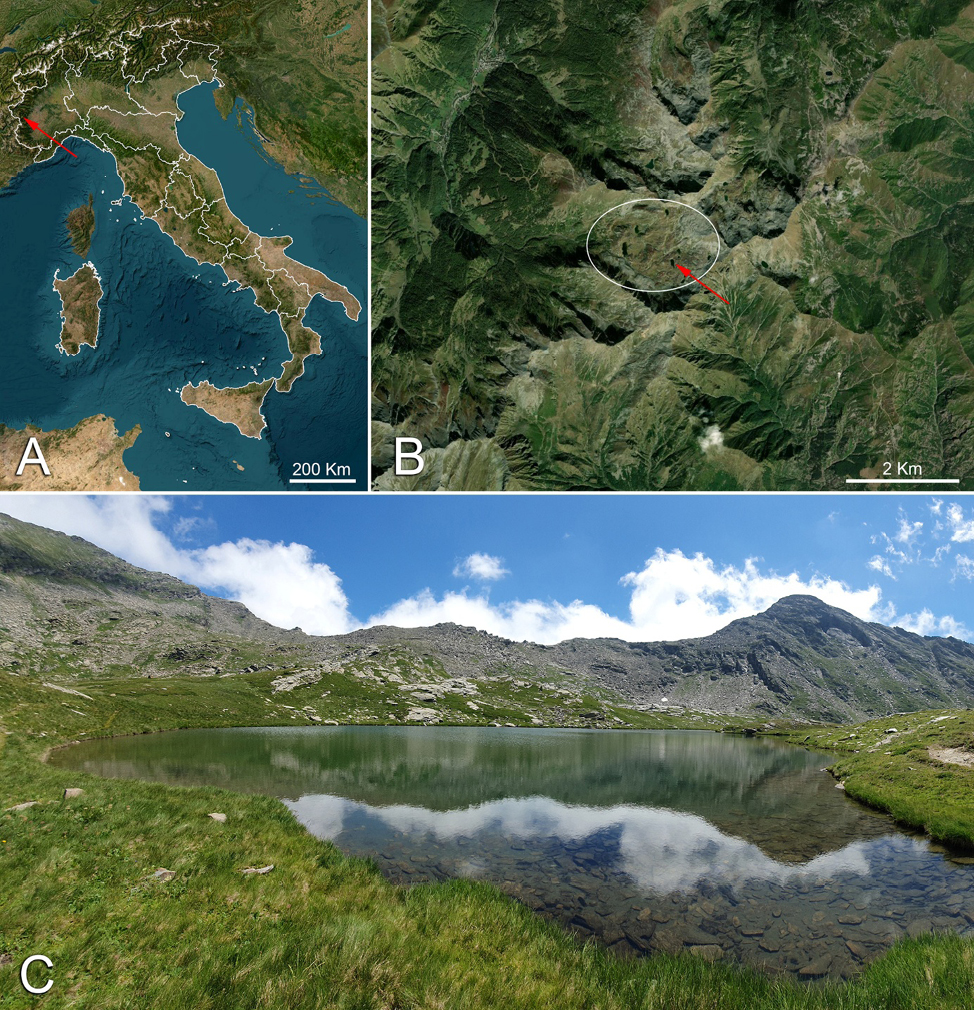
In August 2024, the Istituto Zooprofilattico Sperimentale del Piemonte, Liguria e Valle d’Aosta was alerted to a MME involving Rana temporaria. The event was observed in the waters of “Lago Lungo,” a small alpine lake located within the municipality of Prali (Turin, Piedmont; coordinates: 44.8578, 07.0907; altitude: 2503 m a.s.l.), in the area known as the “Conca dei Tredici Laghi” (hereafter “Conca”, from the Italian for “basin”). This high-altitude basin is a glacial cirque situated in the central sector of the Cottian Alps, within the upper Germanasca Valley (Fig. 1). It hosts a series of small glacial lakes formed by Quaternary glaciation and moraine deposition, representing an example of post-glacial alpine landscape dynamics (see Allasia et al., 2004; Nigrelli, 2005; Farina, 2008; Forno et al., 2011). Only two amphibian species occur in the Conca area: the common frog (R. temporaria) and the Lanza’s salamander (Salamandra lanzai Nascetti, Andreone, Capula and Bullini, 1988), similarly to what has been reported for the nearby Conca Cialancia (Seglie, 2020). The former species reproduces in the lakes within the Conca (Di Nicola, pers. obs.), whereas Lanza’s salamander typically gives birth to fully developed, terrestrial juveniles (Bergò and Andreone, 2002; Di Nicola et al., 2021).
A field survey was carried out in the Conca to assess the reported MME and to collect samples for pathogen screening. Dead common frogs were visually inspected, and a subset of the least decomposed individuals was collected and transported under refrigeration for further analysis.
Field inspection and gross pathology
An initial external examination to assess the general condition of the specimens was performed in situ shortly after sampling, to minimise further post-mortem alterations due to transport. Particular attention was paid to the skin and limbs, which were examined for external lesions and gross abnormalities, such as ulcerations, haemorrhages, necrosis, skin sloughing, oedema, and other deviations from normal appearance. Once in the laboratory, skin samples were collected from multiple body regions and pooled per individual for molecular analysis.
Subsequently, a detailed internal examination of the coelomic cavity and visceral organs was performed. Each specimen was positioned in dorsal recumbency, and a sterile scalpel was used to make a ventral midline laparotomy from the intermandibular region to the cloaca. Major organs were inspected, sampled, and pooled per specimen for molecular analysis. Histopathological examinations were not performed due to the poor post-mortem condition of the carcasses.
Bacteriology
Bacteriological analysis was performed following standardised protocols (Pastorino et al., 2021). In detail, the vitreous humour was aseptically inoculated onto Columbia Agar (CBA) (Liofilchem®, Italy) supplemented with 5% sheep blood, after first swabbing the ocular surface with 70% ethanol. Plates were incubated at 22 ± 2 °C for 72 hours with daily evaluation. Dominant colonies were subcultured on CBA and incubated for an additional 24 hours at 22 ± 2°C. Bacterial identification was performed using matrix-assisted laser desorption/ionization time-of-flight mass spectrometry (MALDI-TOF MS) (Bruker Daltonics Inc., Billerica, MA, USA).
Molecular pathogen screening
Bd and ranaviruses are among the most significant infectious agents currently threatening anuran populations in Europe (Price et al., 2014; Allain and Duffus, 2019; Fisher and Garner, 2020) and were therefore selected for the pathogen screening. Herpesviruses were also included, as they have been indicated as potential emerging pathogens of concern (Franklinos et al., 2018; Origgi et al., 2018; Allain and Duffus, 2019). Molecular analyses were performed on both skin samples and pooled target organs, following genomic DNA extraction using the ReliaPrep™ gDNA Tissue Miniprep System (Promega Corporation, Madison, WI, USA), according to the manufacturer’s protocol.
Bd detection was carried out using a real-time TaqMan PCR protocol targeting the ITS-1/5.8S rDNA region (Table 1; Boyle et al., 2004; Meletiadis et al., 2025). The following thermal conditions were applied: 50°C for 2 min, 95°C for 10 min, followed by 50 cycles of 95°C for 15 s and 60°C for 1 min. Primers and probe used were ITS1-3 Chytr, 5.8S Chytr, and Chytr MGB2, respectively. Genomic DNA from Bd-positive Pelophylax sp. skin was used as a positive control.
| Target gene | Primer | Amplicon size (bp) | Nucleotide sequence (5’ to 3’) | Reference |
|---|---|---|---|---|
| ITS-1/5.8S rDNA | ITS1-3 Chytr | 146 | CCTTGATATAATACAGTGTGCCATATGTC | Boyle et al., 2004 |
| 5.8S Chytr | AGCCAAGAGATCCGTTGTCAAA | |||
| Chytr MGB2 | 6FAM CGAGTCGAACAAAAT MGBNFQ | |||
| DNA pol | DFA (step1 forward) | 725 | GAYTTYGCNAGYYTNTAYCC | Bianchessi et al., 2024 |
| GAYTTYGCIaAGYYTIaTAYCC | ||||
| ILK (step1 forward) | 470 | TCCTGGACAAGCARNYSGCNMTNAA | ||
| TCCTGGACAAGCARIaYSGCIaMTIaAA | ||||
| KG1 (step1 reverse) | GTCTTGCTCACCAGNTCNCANCCYTT | |||
| GTCTTGCTCACCAGIaTCIaCAIaCCYTT | ||||
| TGV | 225 | TGTAACTCGGTGTAYGGNTTYACNGGNGT | ||
| TGTAACTCGGTGTAYGGIaTTYACIaGGIaGT | ||||
| IYG | TGTAACTCGGTGTAYGGIaTTYACIaGGIaGT | |||
| CACAGAGTCCGTRTCIaCCRTAIaAT | ||||
| MCP | OLT-1 | 531 | GACTTGGCCACTTATGAC | Mao et al., 1997 |
| OLT-2R | GTCTCTGGAGAAGAAGAAT |
Herpesvirus detection followed a pan-herpesvirus nested PCR protocol with ten degenerate and deoxyinosine-substituted primers (Table 1 targeting a conserved region of the DNA polymerase gene (Ehlers et al., 1999; Franklinos et al., 2018; Bianchessi et al., 2024). The following thermal conditions were applied for both rounds of the nested PCR: initial activation at 95°C for 15 min, followed by 45 cycles of denaturation at 94°C for 30 s, annealing at 46°C for 60 s, and extension at 72°C for 60 s, with a final elongation at 72°C for 10 min. Genomic DNA from a herpesvirus-positive lemur sample was used as a positive control.
Ranavirus detection was conducted using an endpoint PCR protocol targeting the major capsid protein (MCP) gene, using primers OL T1_F and OL T2_R (Table 1; Mao et al., 1997; Stöhr et al., 2013). The following thermal conditions were applied: activation of Taq polymerase at 95°C for 2 min, followed by 35 cycles of denaturation at 95°C for 1 min; annealing at 50°C for 1 min; extension at 72°C for 1 min; and final elongation at 72°C for 10 min. A synthetic positive control amplicon of approximately 500 bp was included.
All DNA extracts were tested in technical duplicate across assays. No-template controls and extraction blanks were included in every run.
Sequencing and phylogenetic analysis
For Ranavirus-positive samples, Sanger sequencing was performed. Amplification products were checked by electrophoresis on a 2% agarose gel, purified using the ExtractMe DNA Clean-Up kit (Blirt, Gdańsk, Poland), and amplified with the BrilliantDye™ Terminator v3.1 Cycle Sequencing Kit (NimaGen, Nijmegen, The Netherlands). Sequencing reaction products were then purified using the DyeEx 2.0 Spin Kit (Qiagen, Hilden, Germany) and run on a SeqStudio™ Genetic Analyzer (Applied Biosystems, Waltham, MA, USA).
The phylogenetic analysis for ranavirus was performed by comparing the major part of the MCP gene in overlapping fragments (almost complete CDS and partial 3’UTR) and a partial sequence of the DNA polymerase gene (DNApol). The amplification, purification and sequencing were performed as previously described for the diagnostic PCR, using the primers listed in Table 2. All the primers were available in literature, except the reverse primer MCP-6Rb. The alignment between primer and ranavirus sequences showed that the MCP-6R primer developed by Hyatt et al. (2000) for the amplification of the last fragment of the MCP gene was localized in a region characterized by a two-nucleotide polymorphism. To avoid amplification failure, the new primer MCP-6Rb was developed in the 3’UTR region of the gene.
| Target gene | Primer | Primer position | Amplicon size (bp) | Nucleotide sequence (5’ to 3’) | Reference |
|---|---|---|---|---|---|
| MCP | OLT-1 | 97387-97404 | 531 | GACTTGGCCACTTATGAC | Mao et al., 1997 |
| OLT-2R | 97917-97899 | GTCTCTGGAGAAGAAGAAT | |||
| MCP-BF | 97813-97830 | 548 | ACCAGCGATCTCATCAAC | Ariel et al., 2010 | |
| MCP-BR | 98360-98341 | AGGGCTGGCTCCAGGACCGT | |||
| MCP-5 | 98244-98263 | 622 | CGCAGTCAAGGCYTGATGT | Hyatt et al., 2000 | |
| MCP-6Rb | 98865-98843 | TGCCATGTTAAGATTTGTCAGAG | This study | ||
| DNA pol | DNApol-F | 67188-67208 | 560 | GTGTAYCAGTGGTTTTGCGAC | Holopainen et al., 2009 |
| DNApol-R | 67747-67728 | TCGTCTCCGGGYCTGTCTTT |
For each gene, the obtained sequences were aligned with similar regions from 24 complete genomes of the different ranavirus species presented in GenBank (Table S1), using Clustal W program on MEGA7 software. The sequences of MCP and DNA polymerase genes from Lymphocystis disease virus (LCDV) were used as an outgroup. Phylogenetic trees were constructed using MEGA7 software (Kumar et al., 2016) with the maximum likelihood method, using General Time Reversible model, uniform substitution type and invariable sites. All positions containing gaps and missing data were eliminated. The statistical robustness and reliability of the branching order were confirmed with bootstrap analysis using 1000 reiterations.
Results
Field observations and gross clinical findings
During approximately one hour of field inspection, a total of 78 dead common frogs were recorded, all of which were adults except for two subadults. Of these, 73 were found on the bottom of Lago Lungo (generally within three metres of the shoreline; Fig. 2 A, B), three were found desiccated along the shore (Fig. 2 C), and two were observed in the outflow stream descending from the lake. Most carcasses exhibited advanced post-mortem decomposition, with several specimens reduced to skeletal remains. Moreover, limited lakebed visibility (about 5-6 meters from the shoreline) may have led to an underestimation of the total number of dead common frogs.
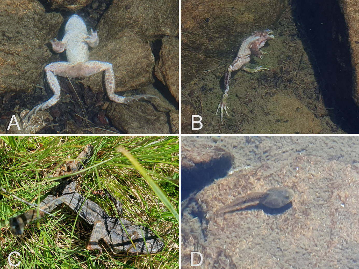
No dead frogs were observed in the other lakes of the Conca. In both Lago Lungo and the adjacent lakes, live adult frogs (n = 9) and tadpoles (n = 4; Fig. 2 D) were present and appeared clinically healthy upon visual inspection, with no external lesions or abnormalities detected.
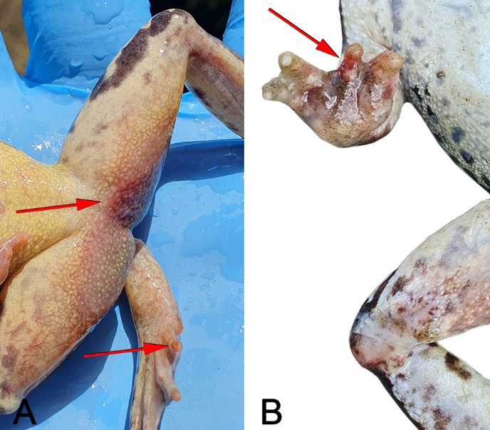
Out of the 78 dead common frogs observed, nine specimens were collected for laboratory analysis; all were adult males. Six of these (specimens I, III, V, VII, VIII and IX) exhibited red discolouration consistent with hyperaemia or haemorrhage, either localised in the ventral thigh region or extending across the entire ventral surface of the hind limbs and/or the digits (Fig. 3). In three of these cases, ulcerative lesions were also present on the toes (I, III and VII). The remaining three frogs (specimens II, IV and VI) did not display any visible external clinical signs, apart from post-mortem alterations. At necropsy, all specimens showed post-mortem alterations, including partial liquefaction of internal organs in some cases, which limited thorough assessment. However, gastrointestinal haemorrhage was observed in three individuals.
Laboratory findings and phylogenetic analysis
Bacteriological cultures were positive in eight out of nine frogs. The isolated bacterial species were: Hafnia alvei (specimens I, IV, and V); Acinetobacter guillouiae (specimens III, VI and VIII); Acinetobacter proteolyticus (specimen IX); and Serratia proteamaculans (specimen VII) (Table 3).
| Specimen | Clinical signs | Ranavirus PCR positive | Hafnia alvei | Acinetobacter guillouiae | Acinetobacter proteolyticus | Serratia proteamaculans |
|---|---|---|---|---|---|---|
| I | + | + | + | - | - | - |
| II | - | - | - | - | - | - |
| III | + | + | - | + | - | - |
| IV | - | - | + | - | - | - |
| V | + | + | + | - | - | - |
| VI | - | + | - | + | - | - |
| VII | + | - | - | - | - | + |
| VIII | + | - | - | + | - | - |
| IX | + | - | - | - | + | - |
All frogs tested negative for Bd and herpesvirus DNA in both skin and pooled target organ samples.Regarding ranavirus, skin samples from four frogs (specimens I, III, V and VI) tested positive using the diagnostic end-point PCR (Table 3). The obtained sequences were 98% identical to common midwife toad virus (CMTV) entries in the NCBI database. Based on this result, a phylogenetic analysis was conducted.
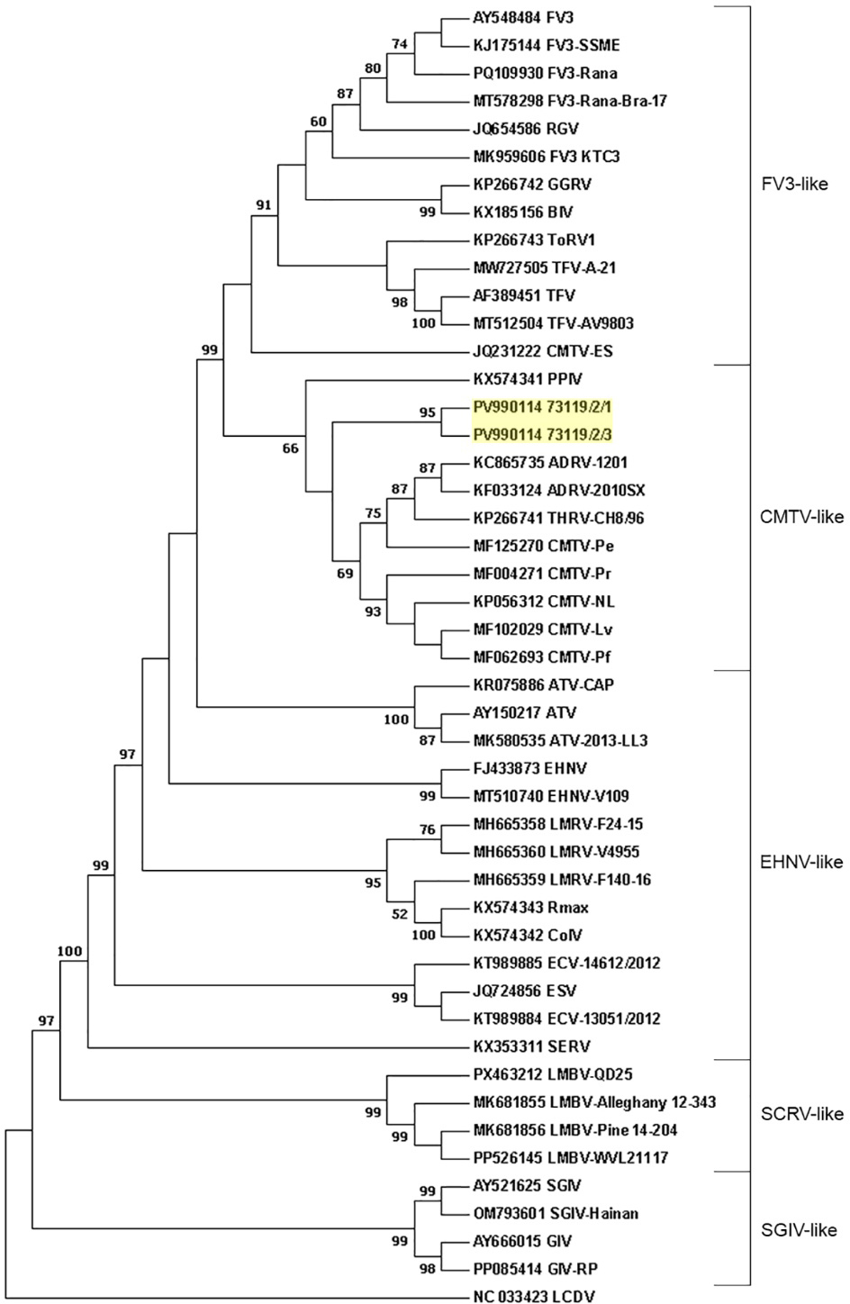
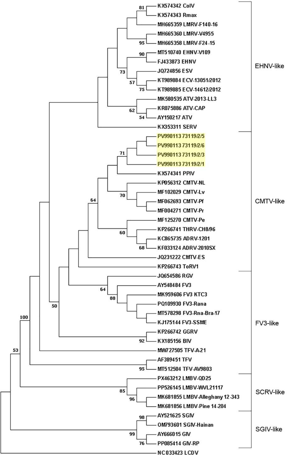
A 1437 bp fragment of the MCP gene was successfully amplified for two samples (specimens I and III), while a 496 bp fragment of the DNA polymerase gene was obtained from all four PCR-positive individuals. The sequences showed no polymorphisms among the samples and the lengths corresponded to the similar CMTV-like sequences deposited in NCBI database. The obtained MCP and DNA polymerase gene sequences were deposited in GenBank under accession numbers PV990114 and PV990113, respectively.
The sequences of both genes were similar to CMTV-like sequences with a percentage over 99%, except for TorV-1 and CMTV-ES which similarity was 98% with our samples. The similarity with other ranaviruses ranged from 97-98 % but decreased below 80% with SCRV (Santee-Cooper ranavirus) and SGIV (Singapore grouper iridovirus)-like groups. This situation was evident in the phylogenetic analysis where the samples clustered with other sequences belonging to the CMTV-like group for both the genes (Fig. 4 and Fig. 5). Considering the MCP gene, CMTV- and FV3-like groups clustered more closely together with high support (99%) in respect to EHNV-like (Epizootic haematopoietic necrosis virus) paraphyletic group, that includes ATV (Ambystoma tigrinum virus), EHNV, ENAR (European North Atlantic ranavirus) and SERV (Short-finned eel ranavirus). The most basally branching lineages were SCRV- and SGIV-like. The DNA polymerase gene sequence analysis showed a similar result (considering that the FV3-like group was paraphyletic, while EHNV-like was monophyletic) but with little support.
Discussion
In the present study, we document the detection of a CMTV-like ranavirus following a summer MME affecting Rana temporaria in a high-altitude lake in the Cottian Alps, Piedmont, Italy. Ranaviral DNA was detected by PCR in four of nine carcasses and confirmed by Sanger sequencing of fragments of the MCP and DNA polymerase genes, with phylogenetic analysis placing these sequences within the CMTV-like clade. Several frogs, both among those tested and among additional carcasses observed in the field, showed clinical signs consistent with ranaviral disease, including ventral red discolouration consistent with hyperaemia/haemorrhage, as well as ulcerative and necrotic lesions affecting the digits.
Bacteriological analysis provided further insights into the condition of the frogs. Bacterial colonies were isolated from the vitreous humour in eight out of nine individuals; since this compartment is anatomically protected, the presence of bacteria is generally interpreted as evidence of systemic infection rather than external contamination (Hanna et al., 1990; Masli and Vega, 2011; Pigaiani et al., 2020). The identified bacteria, including H. alvei, A. guillouiae, A. proteolyticus and S. proteamaculans, are commonly found in the environment or in the gut, but have also been reported as opportunistic pathogens in immunocompromised or environmentally stressed animals (Mahlen, 2011; Padilla et al., 2015; Ionescu et al., 2022; Trinh and Nguyen, 2024). Of particular interest, A. guillouiae has recently been shown to cause systemic disease in amphibians, with experimental infections in Quasipaa spinosa resulting in tissue damage and mortality (Guo et al., 2025). Although the exact contribution of these bacteria to the mortality event cannot be determined, their recovery from an internal immune-privileged site supports the hypothesis of opportunistic invasion, possibly favoured by environmental stressors and host immunosuppression associated with viral infection. Anyway, findings from autolysing amphibian carcasses should be interpreted with caution (White and Dusek, 2015). Although histopathological analyses were not performed, the molecular evidence and observed external lesions support ranavirus infection as the most plausible primary cause of the MME.
To further characterise the outbreak, sequence data from two gene regions (MCP and DNA polymerase) were obtained from ranavirus-positive R. temporaria samples. Both sequences showed high similarity and grouped unequivocally within the CMTV-like group.
Previous phylogeographic studies indicated that the genetic similarity of ranaviruses often reflects their geographic origin rather than host-specific relationships, demonstrating significant differences among European ranavirus strains compared to those from other regions globally (Stöhr et al., 2015). Specifically, CMTV-like ranaviruses are broadly distributed in continental Europe, with only limited detections in Asia and records confined to aquaculture facilities in North America (Claytor et al., 2017; Herath et al., 2023; Lisachova et al., 2025; Marschang et al., 2025). Available evidence indicates that CMTV-like ranaviruses are likely endemic to Europe, with the Iberian Peninsula emerging as a hotspot of genetic diversity. Multilocus analyses from multiple independent outbreaks in Spain over recent decades have identified novel Iberian genotypes and revealed phylogeographic patterns consistent with an ancestral Iberian origin followed by natural dispersal. Nonetheless, the precise origin remains unresolved (Thumsová et al., 2022).
From an evolutionary perspective, CMTV occupies an intermediate position among ranaviruses, retaining features characteristic of EHNV-like viruses but also acquiring unique genomic segments typical of FV3-like viruses (Mavian et al., 2012). The divergence between FV3 and CMTV lineages appears recent, as their separation cannot always be clearly resolved based solely on phylogenetic distance trees (Stöhr et al., 2015). Yu et al. (2021) tried to better examine the classification within ranavirus subgroups by a genomic phylogenetic analysis and a dot plot comparison and identified four genomic regions that consistently matched whole-genome analyses. They excluded the DNA polymerase gene due to recombination events and rejected the MCP gene for occasional inconsistencies despite a lack of recombination or substitution saturation. For example, they highlighted a discrepancy in the phylogenetic tree considering only the MCP gene sequences: the samples TorV1 and CMTV-ES were closely related to the FV3-like group, while they should belong to CMTV-like group based on genome analysis. Consistent with these observations, our phylogenetic analysis based on MCP gene fragments showed CMTV-ES and ToRV-1 grouped with the FV3-like viruses. Conversely, they clustered in the CMTV-like group considering DNA polymerase gene (even if with little support).
Overall, the phylogenetic analyses based on both DNA polymerase and MCP gene sequences conclusively placed our samples within the CMTV-like group, providing the first geolocated detection of CMTV-like ranavirus in free-ranging amphibians in Italy, expanding the documented range of this pathogen and highlighting its potential threat to alpine amphibian populations. The only other documented Italian instance was reported by Holopainen et al. (2009), who isolated Rana esculenta virus 282/I02 (REV 282/I02) from Pelophylax esculentus tadpoles collected from an unspecified locality in 2002 (see Ariel et al., 2017); the larvae developed disease and died shortly after transfer to captivity (Holopainen et al., 2009; Ariel et al., 2010, 2017). Subsequent genomic analyses placed REV 282/I02 within the CMTV-like clade (Ariel et al., 2017). In addition, online outreach articles report that ranavirus has been found in Italian territory, with ongoing investigations in the Maritime Alps in Piedmont (Aree Protette Alpi Marittime, 2019; Piemonte Parchi, 2019), but no peer-reviewed data from these surveys are currently available. Given the geographic proximity to the Mercantour outbreak sites in France (Miaud et al., 2016, 2019), it will be important to document any ranavirus detections that may occur in the Maritime Alps and to assess the phylogenetic placement of any strains involved, particularly with respect to the CMTV-like clade.
CMTV-like ranaviruses have been associated with multiple mortality events in Spain and France, in some cases even contributing to community-level declines (Price et al., 2014, Miaud et al., 2016, Thumsová et al., 2022). In Spain, one of the most recent outbreaks occurred in the northwest, affecting Pleurodeles waltl populations in small cattle ponds that typically dry in summer. Although Bd co-occurred at these sites, coinfection within individuals was rare. Ranavirus loads declined in the months following the outbreak, likely as a result of lower temperatures (Thumsová et al., 2024). By contrast, in the French Alps CMTV caused mass mortality of R. temporaria across several alpine lakes, affecting both larvae and adults, Bd was not detected, and experimental infections produced 100 % mortality in adults (Miaud et al., 2016). Our event shares the alpine setting and late-summer timing, but it was dominated by adult carcasses while tadpoles were observed alive, consistent with a context-dependent expression of CMTV impacts across systems.
Our samples were also screened for Bd and herpesviruses, with no positive detections. Although Bd was not detected at our site, Bd and ranaviruses can co-occur at the same localities, whereas coinfection within individuals is usually rare and infection with one pathogen does not reliably predict infection with the other (Bosch et al., 2020; Thumsová et al., 2024). This pattern is consistent with evidence that the two pathogens tend to peak under contrasting temperature and precipitation conditions, which may limit temporal overlap even where they co-occur geographically (Thumsová et al., 2025).
Anthropogenic activities such as wildlife trade and introduction of non-native species can facilitate ranavirus spread into novel ecosystems and host communities. In particular, fish introduced through aquaculture or stocking have been linked with amphibian outbreaks, and invasive fish can also disrupt pre-existing host-pathogen equilibria, increasing ranavirus prevalence and driving long-term declines even without introducing novel strains (Price et al., 2017; Rosa et al., 2022). In line with this, targeted screening of non-native fish species should be included in future monitoring at our study site, as they are present in the Conca for recreational fishing and may contribute to local transmission or amplification (Peñafiel-Ricaurte et al., 2025). In addition, environmental DNA-based surveillance from water samples may increase ranavirus detectability and support non-invasive monitoring, even when no overt die-off is observed (Miaud et al., 2019).
Although the drivers of ranavirus emergence in our study system remain unclear, climatic variability has consistently been linked to ranavirus dynamics in other regions. Warmer conditions have been associated with increased incidence and severity of outbreaks, and several studies have related ranavirus epizootics to ongoing global warming (Price et al., 2019; Thumsová et al., 2022). More recently, a large-scale study from the Iberian Peninsula suggested that increased ranavirus infection risk is not driven by rising temperatures alone, but rather by mismatches between local host adaptations and complex interactions between changing temperature and precipitation patterns (Thumsová et al., 2025). Accordingly, future work should integrate continuous water temperature logging, local precipitation metrics and basic physicochemical profiling (for example dissolved oxygen, pH, conductivity), and consider microhabitat features such as shading to better characterise conditions that may favour pathogen emergence. The surveyed area also hosts Salamandra lanzai, a live-bearing species endemic to the Cottian Alps and classified as “Vulnerable” in the Italian IUCN Red List of vertebrates (Rondinini et al., 2022); its inclusion in ranavirus surveillance programmes is advisable, given previous detections of ranavirus within the genus Salamandra (Vörös et al., 2020).
In summary, despite the limited number of specimens analysed and the lack of histopathology, our survey confirms the presence of a CMTV-like ranavirus in common frogs within the Italian Alpine range and provides the first geolocated detection of this lineage in free-ranging amphibians in Italy. These findings call for more detailed investigations to better characterise ranavirus circulation and clarify its role in mortality events, integrating host screening across taxa with targeted environmental monitoring.
Acknowledgements
The authors thank Andrea Samorè and Giulia Tessa. The sample collection was carried out under the following permits: MiTE/Ministry of Ecological Transition (MiTE N. 0024526.28-02-2022), approved by ISPRA on 23 February 2022 (Protocol N. 0009384/2022).
Supplementary material
Supplementary material associated with this article can be found at https://oaj.fupress.net/index.php/ah/article/view/18382/15289.
References
Akçakaya, H.R., Neam, K., Hobin, L., Lötters, S., Martel, A., Pasmans, F. (2023): Assessing the extinction risks of amphibians impacted by infectious diseases. Biol. Conserv. 284: 110205.
Allain, S.J., Duffus, A.L. (2019): Emerging infectious disease threats to European herpetofauna. Herpetol. J. 29: 189-206.
Allasia, P., Audisio, C., Baldo, M., Cirio, C.G., Lollino, G., Giordan, D., Godone, F., Nigrelli, G., Alpe, F., Ambrogio, S., Giardino, M., Perotti, L., Sambuelli, L., De Renzo, G., Fontan, D., Barbero, T. (2004): Instrumented experimental sites for the control of landslide hazards in mountain environments: the Germanasca and Susa Valleys (Northwestern Italy). In: Field trip guidebooks, 32nd International Geological Congress, Florence, Italy, 20-28 August 2004, pp. 37-54. APAT, Roma.
Aree Protette Alpi Marittime (2019): Ranavirus a rischio la rana temporaria. Aree Protette Alpi Marittime.
Ariel, E., Holopainen, R., Olesen, N.J., Tapiovaara, H. (2010): Comparative study of ranavirus isolates from cod (Gadus morhua) and turbot (Psetta maxima) with reference to other ranaviruses. Arch. Virol. 155: 1261-1271.
Ariel, E., Subramaniam, K., Imnoi, K., Sriwanayos, P., Ahasan, M.S., Olesen, N.J., Amedeo, M., Toffan, A., Waltzek, T.B. (2017): Genomic sequencing of Ranaviruses isolated from edible frogs (Pelophylax esculentus). Genome Announc. 5: 10-1128.
Balseiro, A., Dalton, K., Del Cerro, A., Marquez, I., Cunningham, A., Parra, F., Prieto, J., Casais, R. (2009): Pathology, isolation and molecular characterisation of a Ranavirus from the common midwife toad Alytes obstetricans on the Iberian Peninsula. Dis. Aquat. Org. 84: 95-104.
Bates, K.A., Rosa, G.M., Gamer, T.W.J. (2025): Ranavirus. Trends Microbiol. 33: 701-702.
Bergò, P.E., Andreone, F. (2002): A terrestrial viviparous salamander into water: Notes on the unusual larval aquatic development in Salamandra lanzai. Herpetol. Bull. 81: 29-31.
Bianchessi, L., Flach, E., Monacchia, G., Dagleish, M., Maley, M., Turin, L., Rocchi, M.S. (2024): Identification and characterisation of gamma-herpesviruses in zoo artiodactyla. Virol. J. 21: 1-15.
Bison, M., Yoccoz, N.G., Carlson, B.Z., Klein, G., Laigle, I., Van Reeth, C., Delestrade, A. (2021): Earlier snowmelt advances breeding phenology of the common frog (Rana temporaria) but increases the risk of frost exposure and wetland drying. Front. Ecol. Evol. 9: 1-12.
Bosch, J., Carrascal, L.M., Durán, L., Walker, S., Fisher, M.C. (2007): Climate change and outbreaks of amphibian chytridiomycosis in a montane area of Central Spain: Is there a link? Proc. R. Soc. B. 274: 253-260.
Bosch, J., Monsalve-Carcaño, C., Price, S.J., Bielby, J. (2020): Single infection with Batrachochytrium dendrobatidis or Ranavirus does not increase probability of co-infection in a montane community of amphibians. Sci. Rep. 10: 21115.
Bosch, J., Mora-Cabello De Alba, A., Marquínez, S., Price, S.J., Thumsová, B., Bielby, J. (2021): Long-term monitoring of amphibian populations of a national park in northern Spain reveals negative persisting effects of Ranavirus, but not Batrachochytrium dendrobatidis. Front. Vet. Sci. 8: 645491.
Box, E.K., Cleveland, C.A., Subramaniam, K., Waltzek, T.B., Yabsley, M.J. (2021): Molecular confirmation of Ranavirus infection in amphibians from Chad, Africa. Front. Vet. Sci. 8: 733939.
Boyle, D., Boyle, D., Olsen, V., Morgan, J., Hyatt, A. (2004): Rapid quantitative detection of chytridiomycosis (Batrachochytrium dendrobatidis) in amphibian samples using real-time Taqman PCR assay. Dis. Aquat. Org. 60: 141-148.
Brunner, J.L., Olson, D.H., Gray, M.J., Miller, D.L., Duffus, A.L.J. (2021): Global patterns of Ranavirus detections. FACETS 6: 912-924.
Catalan, J., Ninot, J.M., Aniz, M.M. (2017): High mountain conservation in a changing world. Cham, Springer International Publishing.
Claytor, S.C., Subramaniam, K., Landrau-Giovannetti, N., Chinchar, V.G., Gray, M.J., Miller, D.L., Mavian, C., Salemi, M., Wisely, S., Waltzek, T.B. (2017): Ranavirus phylogenomics: Signatures of recombination and inversions among bullfrog ranaculture isolates. Virology 511: 330-343.
Di Nicola, M.R., Cavigioli, L., Luiselli, L., Andreone, F. (2021): Anfibi & Rettili d’Italia. Edizione aggiornata. Latina; Edizioni Belvedere.
Di Nicola, M.R., Rubiola, S., Cerullo, A., Basciu, A., Massone, C., Zabbia, T., Dorne, J.L.M.C., Acutis, P.L., Marini, D. (2025): Microorganisms in wild European reptiles: Bridging gaps in neglected conditions to inform disease ecology research. Int. J. Parasitol. Parasites. Wildl. 27: 101113.
Duffus, A.L., Nichols, R.A., Garner, T.W. (2014): Detection of a frog virus 3-like Ranavirus in native and introduced amphibians in the United Kingdom in 2007 and 2008. Herpetol. Rev. 45: 608-610.
Ehlers, B., Borchers, K., Grund, C., Frölich, K., Ludwig, H., Buhk, H.-J. (1999): Detection of new DNA polymerase genes of known and potentially novel herpesviruses by PCR with degenerate and deoxyinosine-substituted primers. Virus Genes 18: 211-220.
Farina, T., Ed. (2008): Alpi Cozie. Supplemento a Piemonte Parchi 5(XXIII). Torino, Regione Piemonte.
Fisher, M.C., Garner, T.W.J. (2020): Chytrid fungi and global amphibian declines. Nat. Rev. Microbiol. 18: 332-343.
Fisher, M.C., Henk, D.A., Briggs, C.J., Brownstein, J.S., Madoff, L.C., McCraw, S.L., Gurr, S.J. (2012): Emerging fungal threats to animal, plant and ecosystem health. Nature 484: 186-194.
Flechas, S., Urbina, J., Crawford, A., Gutiérrez, K., Corrales, K., Castellanos, L., González, M., Cuervo, A., Catenazzi, A. (2023): First evidence of ranavirus in native and invasive amphibians in Colombia. Dis. Aquat. Org. 153: 51-58.
Forno, M.G., Lingua, A., Lo Russo, S., Taddia, G. (2011): Improving digital tools for Quaternary field survey: A case study of the Rodoretto Valley (NW Italy). Environ. Earth. Sci. 64: 1487-1495.
Forzán, M.J., Jones, K.M., Ariel, E., Whittington, R.J., Wood, J., Markham, R.J.F., Daoust, P.-Y. (2017): Pathogenesis of frog virus 3 (Ranavirus, Iridoviridae) infection in wood frogs (Rana sylvatica). Vet. Pathol. 54: 531-548.
Franklinos, L., Fernandez, J., Hydeskov, H., Hopkins, K., Everest, D., Cunningham, A., Lawson, B. (2018): Herpesvirus skin disease in free-living common frogs Rana temporaria in Great Britain. Dis. Aquat. Org. 129: 239-244.
Guo, L., Jin, X., Yang, D., Wei, L., Chen, J., Lin, Z., Ma, L. (2025): Identification and characterization of Serratia nematophila and Acinetobacter guillouiae from putrid-skin disease lesions in farmed Chinese spiny frog (Quasipaa spinosa). Microbiol. Spectr. 13: e02096-24.
Hanna, P.E., Bellamy, J.E., Donald, A. (1990): Postmortem eyefluid analysis in dogs, cats and cattle as an estimate of antemortem serum chemistry profiles. Can. J. Vet. Res. 54: 487-494.
Hartmann, A.M., Maddox, M.L., Ossiboff, R.J., Longo, A.V. (2022): Sustained Ranavirus outbreak causes mass mortality and morbidity of imperiled amphibians in Florida. EcoHealth 19: 8-14.
Hick, P.M., Subramaniam, K., Thompson, P., Whittington, R.J., Waltzek, T.B. (2016): Complete genome sequence of a Bohle iridovirus isolate from ornate burrowing frogs (Limnodynastes ornatus) in Australia. Genome Announc. 4: e00632-16.
Herath, J., Sun, D., Ellepola, G., Subramaniam, K., Meegaskumbura, M. (2023): Emerging threat of Ranavirus: Prevalence, genetic diversity, and climatic drivers of Ranavirus (Iridoviridae) in ectothermic vertebrates of Asia. Front. Vet. Sci. 10: 1291872.
Hoberg, E.P., Brooks, D.R. (2015): Evolution in action: Climate change, biodiversity dynamics and emerging infectious disease. Philos. Trans. R. Soc. B 370: 20130553.
Holopainen, R., Ohlemeyer, S., Schütze, H., Bergmann, S., Tapiovaara, H. (2009): Ranavirus phylogeny and differentiation based on major capsid protein, DNA polymerase and neurofilament triplet H1-like protein genes. Dis. Aquat. Org. 85: 81-91.
Hyatt, A.D., Gould, A.R., Zupanovic, Z., Cunningham, A.A., Hengstberger, S., Whittington, R.J., Kattenbelt, J., Coupar, B.E.H. (2000): Comparative studies of piscine and amphibian iridoviruses. Arch. Virol. 145: 301-331.
Ilić, M., Bugarski-Stanojević, V., Jovanović, B., Stamenković, G., Zorić, K., Paunović, M., Crnobrnja-Isailović, J. (2024): Phylogeographic substructuring in the southernmost refugium of the European common frog Rana temporaria. Animals 14: 1430.
Ionescu, M.I., Neagoe, D. Ștefan, Crăciun, A.M., Moldovan, O.T. (2022): The gram-negative bacilli isolated from caves – Sphingomonas paucimobilis and Hafnia alvei and a review of their involvement in human infections. Int. J. Environ. Res. Public Health 19: 2324.
Jancovich, J.K., Steckler, N.K., Waltzek, T.B. (2015): Ranavirus taxonomy and phylogeny. In: Ranaviruses; pp. 59-70. Gray, M.J., Chinchar, V.G., Eds, Cham, Springer International Publishing.
Knapp, R.A., Briggs, C.J., Smith, T.C., Maurer, J.R. (2011): Nowhere to hide: Impact of a temperature-sensitive amphibian pathogen along an elevation gradient in the temperate zone. Ecosphere 2: art93.
Kumar, S., Stecher, G., Tamura, K. (2016): MEGA7: Molecular Evolutionary Genetics Analysis version 7.0 for Bigger Datasets. Mol. Biol. Evol. 33: 1870-1874.
Lisachova, L.S., Lisachov, A.P., Ermakov, O.A., Svinin, A.O., Chernigova, P.I., Lyapkov, S.M., Zamaletdinov, R.I., Pavlov, A.V., Zaks, S.S., Fayzulin, A.I., Korzikov, V.A., Simonov, E. (2025): Continent-wide distribution of CMTV-like Ranavirus, from the Urals to the Atlantic Ocean. EcoHealth 22: 173-184.
Luedtke, J.A., Chanson, J., Neam, K., Hobin, L., Maciel, A.O., Catenazzi, A., Borzée, A., Hamidy, A., Aowphol, A., Jean, A., Sosa-Bartuano, Á., Fong G., A., De Silva, A., Fouquet, A., Angulo, A., Kidov, A.A., Muñoz Saravia, A., Diesmos, A.C., Tominaga, A., Shrestha, B., Gratwicke, B., Tjaturadi, B., Martínez Rivera, C.C., Vásquez Almazán, C.R., Señaris, C., Chandramouli, S.R., Strüssmann, C., Cortez Fernández, C.F., Azat, C., Hoskin, C.J., Hilton-Taylor, C., Whyte, D.L., Gower, D.J., Olson, D.H., Cisneros-Heredia, D.F., Santana, D.J., Nagombi, E., Najafi-Majd, E., Quah, E.S.H., Bolaños, F., Xie, F., Brusquetti, F., Álvarez, F.S., Andreone, F., Glaw, F., Castañeda, F.E., Kraus, F., Parra-Olea, G., Chaves, G., Medina-Rangel, G.F., González-Durán, G., Ortega-Andrade, H.M., Machado, I.F., Das, I., Dias, I.R., Urbina-Cardona, J.N., Crnobrnja-Isailović, J., Yang, J.-H., Jianping, J., Wangyal, J.T., Rowley, J.J.L., Measey, J., Vasudevan, K., Chan, K.O., Gururaja, K.V., Ovaska, K., Warr, L.C., Canseco-Márquez, L., Toledo, L.F., Díaz, L.M., Khan, M.M.H., Meegaskumbura, M., Acevedo, M.E., Napoli, M.F., Ponce, M.A., Vaira, M., Lampo, M., Yánez-Muñoz, M.H., Scherz, M.D., Rödel, M.-O., Matsui, M., Fildor, M., Kusrini, M.D., Ahmed, M.F., Rais, M., Kouamé, N.G., García, N., Gonwouo, N.L., Burrowes, P.A., Imbun, P.Y., Wagner, P., Kok, P.J.R., Joglar, R.L., Auguste, R.J., Brandão, R.A., Ibáñez, R., Von May, R., Hedges, S.B., Biju, S.D., Ganesh, S.R., Wren, S., Das, S., Flechas, S.V., Ashpole, S.L., Robleto-Hernández, S.J., Loader, S.P., Incháustegui, S.J., Garg, S., Phimmachak, S., Richards, S.J., Slimani, T., Osborne-Naikatini, T., Abreu-Jardim, T.P.F., Condez, T.H., De Carvalho, T.R., Cutajar, T.P., Pierson, T.W., Nguyen, T.Q., Kaya, U., Yuan, Z., Long, B., Langhammer, P., Stuart, S.N. (2023): Ongoing declines for the world’s amphibians in the face of emerging threats. Nature 622: 308-314.
Machate, O., Schmeller, D.S., Schulze, T., Brack, W. (2023): Review: Mountain lakes as freshwater resources at risk from chemical pollution. Environ. Sci. Eur. 35: 3.
Mahlen, S.D. (2011): Serratia infections: From military experiments to current practice. Clin Microbiol. Rev. 24: 755-791.
Mao, J., Hedrick, R.P., Chinchar, V.G. (1997): Molecular characterization, sequence analysis, and taxonomic position of newly isolated fish iridoviruses. Virology 229: 212-220.
Marschang, R.E., Meddings, J.I., Waltzek, T.B., Hick, P., Allender, M.C., Wirth, W., Duffus, A.L.J. (2025): Ranavirus distribution and host range. In: Ranaviruses; pp. 155-230. Gray, M.J., Chinchar, V.G., Eds, Cham, Springer.
Masli, S., Vega, J.L. (2011): Ocular immune privilege sites. Methods Mol. Biol. 677: 449-458.
Mavian, C., López-Bueno, A., Balseiro, A., Casais, R., Alcamí, A., Alejo, A. (2012): The genome sequence of the emerging common midwife toad virus identifies an evolutionary intermediate within ranaviruses. J. Virol. 86: 3617-3625.
Meletiadis, A., Di Nicola, M.R., Bovero, S., Favelli, M., Pezzolato, M., Grella, S., Rezza, G., Acutis, P.L. (2025): Prevalence of Batrachochytrium dendrobatidis in amphibians in northwestern Italy’s protected areas. Animals 15: 157.
Miaud, C., Arnal, V., Poulain, M., Valentini, A., Dejean, T. (2019): eDNA increases the detectability of Ranavirus infection in an alpine amphibian population. Viruses 11: 526.
Miaud, C., Pozet, F., Gaudin, N.C.G., Martel, A., Pasmans, F., Labrut, S. (2016): Ranavirus causes mass die-offs of alpine amphibians in the southwestern alps, France. J. Wildl. Dis. 52: 242-252.
Miller, D.L., Pessier, A.P., Hick, P., Whittington, R.J., Forzán, M.J. (2025): Pathology and diagnostics. In: Ranaviruses; pp. 271-312. Gray, M.J., Chinchar, V.G., Eds, Cham, Springer.
Nigrelli, G. (2005): Analysis and characteristics of pluviometric events in the Germanasca Valley (Italian Western Alps). Geogr. Fis. Din. Quat. 28: 147-158.
North, A.C., Hodgson, D.J., Price, S.J., Griffiths, A.G.F. (2015): Anthropogenic and ecological drivers of amphibian disease (Ranavirosis). PLoS ONE 10: e0127037.
Origgi, F.C., Schmidt, B.R., Lohmann, P., Otten, P., Meier, R.K., Pisano, S.R.R., Moore-Jones, G., Tecilla, M., Sattler, U., Wahli, T., Gaschen, V., Stoffel, M.H. (2018): Bufonid herpesvirus 1 (BfHV1) associated dermatitis and mortality in free ranging common toads (Bufo bufo) in Switzerland. Sci. Rep. 8: 14737.
Padilla, D., Acosta, F., Ramos-Vivas, J., Grasso, V., Bravo, J., El Aamri, F., Real, F. (2015): The pathogen Hafnia alveiin veterinary medicine: A review. J. Appl. Anim. Res. 43: 231-235.
Pastorino, P., Barceló, D., Prearo, M. (2024): Alps at risk: High-mountain lakes as reservoirs of persistent and emerging contaminants. J. Contam. Hydrol. 264: 104361.
Pastorino, P., Colussi, S., Pizzul, E., Varello, K., Menconi, V., Mugetti, D., Tomasoni, M., Esposito, G., Bertoli, M., Bozzetta, E., Dondo, A., Acutis, P.L., Prearo, M. (2021): The unusual isolation of carnobacteria in eyes of healthy salmonids in high-mountain lakes. Sci. Rep. 11: 2314.
Pastorino, P., Prearo, M. (2020): High-mountain lakes, indicators of global change: Ecological characterization and environmental pressures. Diversity 12: 260.
Peñafiel-Ricaurte, A., Price, S.J., Fisher, M.C., Bosch, J., Cunningham, A.A., Azat, C. (2025): Ranavirus in native amphibians from the Pyrenees National Park, France: A case study. Eur. J. Wildl. Res. 71: 39.
Pigaiani, N., Bertaso, A., De Palo, E.F., Bortolotti, F., Tagliaro, F. (2020): Vitreous humor endogenous compounds analysis for post-mortem forensic investigation. Forensic Sci. Int. 310: 110235.
Piemonte Parchi (2019): Ranavirus minaccia la rana temporaria. Piemonte Parchi.
Price, S.J., Ariel, E., Maclaine, A., Rosa, G.M., Gray, M.J., Brunner, J.L., Garner, T.W.J. (2017): From fish to frogs and beyond: Impact and host range of emergent ranaviruses. Virology 511: 272-279.
Price, S.J., Garner, T.W.J., Nichols, R.A., Balloux, F., Ayres, C., Mora-Cabello de Alba, A., Bosch, J. (2014): Collapse of amphibian communities due to an introduced Ranavirus. Curr. Biol. 24: 2586-2591.
Price, S.J., Leung, W.T.M., Owen, C.J., Puschendorf, R., Sergeant, C., Cunningham, A.A., Balloux, F., Garner, T.W.J., Nichols, R.A. (2019): Effects of historic and projected climate change on the range and impacts of an emerging wildlife disease. Glob. Change Biol. 25: 2648-2660.
Råman Vinnå, L., Medhaug, I., Schmid, M., Bouffard, D. (2021): The vulnerability of lakes to climate change along an altitudinal gradient. Commun. Earth. Environ. 2: 35.
Rondinini, C., Battistoni, A., Teofili, C. (2022): Lista Rossa IUCN dei vertebrati italiani 2022. Roma, Comitato Italiano IUCN e Ministero dell’Ambiente e della Sicurezza Energetica.
Rosa, G.M., Sabino-Pinto, J., Laurentino, T.G., Martel, A., Pasmans, F., Rebelo, R., Griffiths, R.A., Stöhr, A.C., Marschang, R.E., Price, S.J., Garner, T.W.J., Bosch, J. (2017): Impact of asynchronous emergence of two lethal pathogens on amphibian assemblages. Sci. Rep. 7: 43260.
Rosa, G.M., Ayala Botto, G., Mitra, A.T., Almeida, J.S.D., Hofmann, M., Leung, W.T.M., Alves De Matos, A.P., Caeiro, M.F., Froufe, E., Loureiro, A., Price, S.J., Owen, C., Rebelo, R., Soares, C. (2022): Invasive fish disrupt host-pathogen dynamics leading to amphibian declines. Biol. Conserv. 276: 109785.
Ruggeri, J., Ribeiro, L.P., Pontes, M.R., Toffolo, C., Candido, M., Carriero, M.M., Zanella, N., Sousa, R.L.M., Toledo, L.F. (2019): Discovery of wild amphibians infected with Ranavirus in Brazil. J. Wildl. Dis. 55: 897-902.
Schilliger, L., Paillusseau, C., François, C., Bonwitt, J. (2023): Major emerging fungal diseases of reptiles and amphibians. Pathogens 12: 429.
Seglie, D. (2020): L’erpetofauna del Parco Naturale di Conca Cialancia, con una nota sulla colorazione atipica di due individui di Salamandra lanzai. Riv. Piem. Stor. Nat. 41: 157-166.
Smith, K.F., Acevedo-Whitehouse, K., Pedersen, A.B. (2009): The role of infectious diseases in biological conservation. Anim. Conserv. 12: 1-12.
Smith, K.F., Sax, D.F., Lafferty, K.D. (2006): Evidence for the role of infectious disease in species extinction and endangerment. Conserv. Biol. 20: 1349-1357.
Stöhr, A.C., Hoffmann, A., Papp, T., Robert, N., Pruvost, N.B.M., Reyer, H.-U., Marschang, R.E. (2013): Long-term study of an infection with ranaviruses in a group of edible frogs (Pelophylax kl. esculentus) and partial characterization of two viruses based on four genomic regions. Vet. J. 197: 238-244.
Stöhr, A.C., López-Bueno, A., Blahak, S., Caeiro, M.F., Rosa, G.M., Alves De Matos, A.P., Martel, A., Alejo, A., Marschang, R.E. (2015): Phylogeny and differentiation of reptilian and amphibian ranaviruses detected in Europe. PLoS ONE 10: e0118633.
Teacher, A.G.F., Cunningham, A.A., Garner, T.W.J. (2010): Assessing the long‐term impact of Ranavirus infection in wild common frog populations. Anim. Conserv. 13: 514-522.
Thumsová, B., Alarcos, G., Ayres, C., Rosa, G.M., Bosch, J. (2024): Relationship between two pathogens in an amphibian community that experienced mass mortalities. Conserv. Biol. 38: e14196.
Thumsová, B., Martínez-Silvestre, A., Pérez-Vallejo, M., Palomar, G., Rohr, J.R., Bosch, J. (2025): Temperature and precipitation mismatches increase infection risk in amphibians. Curr. Biol. 35: 4285-4292.
Thumsová, B., Price, S.J., González-Cascón, V., Vörös, J., Martínez-Silvestre, A., Rosa, G.M., Machordom, A., Bosch, J. (2022): Climate warming triggers the emergence of native viruses in Iberian amphibians. iScience 25: 105541.
Tiberti, R., Rogora, M., Tartari, G., Callieri, C. (2014b): Ecological impact of transhumance on the trophic state of alpine lakes in Gran Paradiso National Park. Knowl. Managt. Aquat. Ecosyst. 415: 05.
Tiberti, R., Von Hardenberg, A. (2012): Impact of introduced fish on Common frog (Rana temporaria) close to its altitudinal limit in alpine lakes. Amphib.-Reptil. 33: 303-307.
Tiberti, R., Von Hardenberg, A., Bogliani, G. (2014a): Ecological impact of introduced fish in high altitude lakes: A case of study from the European Alps. Hydrobiologia 724: 1-19.
Trinh, L.L., Nguyen, H.H. (2024): Role of plant-associated microbes in plant health and development: The case of the Serratia genus. Technol. Agron. 4: e028.
Vörös, J., Herczeg, D., Papp, T., Monsalve-Carcaño, C., Bosch, J. (2020): First detection of Ranavirus infection in amphibians in Hungary. Herpetol. Notes 13: 213-217.
White, C.L., Dusek, R.J. (2015): Wildlife specimen collection, preservation, and shipment. U.S. Geological survey techniques and methods, book 15, chap. C4, 24 p.
Yu, Z., Zhang, W., Gu, C., Chen, J., Zhao, M., Fu, L., Han, J., He, M., Xiao, Q., Xiao, W., He, L., Zhang, Z. (2021): Genomic analysis of Ranavirus and exploring alternative genes for phylogenetics. Transbound. Emerg. Dis. 68: 2161-2170.