https://doi.org/10.36253/a_h-18605
Submitted on: 2025, 9th September; revised on: 2026, 1th April; accepted on: 2026, 10th April.
Life history of the Anji salamander (Chordata, Amphibia, Caudata, Hynobiidae) from fertilized eggs to subadults
1 Department of Basic Medical Science, Huzhou University, 759 Erhuan East Road, Huzhou, Zhejiang 313000, P.R. China
2 Anji Salamander (Hynobius amjiensis) National Nature Reserve Management Center, No. 77-83, Building 1, Yongfu Commercial Street, Baofu Town, Anji County, Huzhou, Zhejiang 313000, P.R. China
3 National-local Joint Engineering Laboratory of Aquatic Animal Genetic Breeding and Nutrition (Zhejiang), Zhejiang Provincial Key Laboratory of Aquatic Resources Conservation and Development, Key Laboratory of Aquatic Animal Genetic Breeding and Nutrition of Chinese Academy of Fishery Sciences, Huzhou University, 759 Erhuan East Road, Huzhou, Zhejiang 313000, P. R. China
*Corresponding authors. Email: 02196@zjhu.edu.cn
Abstract. Anji salamander (Hynobius amjiensis) is a rare species endemic to China, living in peat moss swamp at an altitude of 1,300 meters on Longwang Mountain in Anji County, Zhejiang Province. Anji salamander larvae live in natural puddles in the swamp, and the adults spend most of their time in the humus under the peat moss, only briefly visiting the puddles during the breeding period to spawn and then disappear back into the swamp. Therefore, the life history of Anji salamander is poorly understood. In this study, we analyzed the developmental characteristics, behaviors and life habits of Anji salamander embryos from fertilized eggs to subadults. Following previous studies on the development of small salamanders and amphibians, we divided Anji salamander development into six stages, including embryonic development, balance bar, limb development, pre-metamorphosis, metamorphosis, and subadult development, with 34 developmental nodes. The period before the shedding of the balance bar was a relatively sensitive period. The metamorphosis period, especially the late metamorphosis period, was the most sensitive period from larvae to subadults, during which the small salamanders undergo complex physiological and biochemical changes such as relatively long fasting time, respiratory changes, and transition from aquatic to terrestrial life. These data fill in some of the gaps in the life history of Anji salamander from fertilized eggs to subadults, and are valuable for its conservation and future endeavor on artificial breeding.
Keywords. Anji salamander, Hynobius amjiensis, life history, metamorphosis, molt, subadult.
Contents
Embryo incubation and animal breeding
Fertilized egg and embryonic stages of Anji salamander
Anji salamander balance bar stage
Anji salamander limb developmental stage
Anji salamander pre-metamorphosis stage
Anji salamander metamorphosis stage
Anji salamander subadult stage
General characteristics of the development from fertilized egg to subadult stage of Anji salamander
Embryonic development of Anji salamander
The balance bar of Anji salamander
Patterns of limb development in the Anji salamander
Risks faced during metamorphosis and development of Anji salamander
Introduction
Anji salamander, first discovered in 1991 in Longwang Mountain, Anji County, Zhejiang Province (Gu et al., 1999), and subsequently named Hynobius amjiensis, belongs to the class Amphibia, order Caudata, family Hynobiidae, genus Hynobius. It is a critically endangered (CR) species in China, one of only two families of Hynobiidae in Zhejiang Province (Yang et al., 2016). Previous studies showed that Anji salamander lives in peat moss swamps at an altitude of about 1300 m (Zhang et al., 2010a), and only enters the swamp puddles to spawn during the breeding season from late November to March each year (Liu et al., 2016). Anji salamander lives in peat moss swamps during the non-breeding season. This cryptic habitat preference contributed to its recent discovery (Zhang et al., 2010a). Anji salamander has strict requirements for the quality of its living environment, and the scarcity of its spawning sites and intraspecific competition have led to a very low survival rate of Anji salamander larvae. Therefore, fewer than 1% of hatchlings typically survive to the subadult stange and successfully emerge onto land (Gu et al., 1999; Liu et al., 2016). In recent years, with global warming, the habitat suitable for its survival has been decreasing (Pan et al., 2020). We work in the reserve to explore ways to improve the survival rate through artificial breeding of hatchlings into subadults and then release them back to the land (Chen et al., 2018a). During this process, we also carried out a study on the life history of Anji salamander in order to provide valuable reference data for subsequent conservation works.
Life history refers to the adaptive evolution of morphological, physiological, and behavioral patterns under long-term natural selection in which animals develop their own unique life responses based on their genetic characteristics as well as environmental pressures (Williams, 1966; Begon et al., 1990; Nettle et al., 2019). Life history traits generally involve biological characteristics such as growth, development, and reproduction, and are specifically described as growth patterns, body size, age at sexual maturity, longevity, and clutch size (Stearns, 1977). Amphibians are more vulnerable to habitat loss and fragmentation than other terrestrial animal groups, due to their specialized amphibious life history, relatively weak terrestrial migration, and high dependence on habitat (Tan et al., 2023). With environmental degradation such as reduced precipitation, diseases, and habitat loss, amphibians are facing tremendous survival challenges. The study of internal and external factors such as amphibian life history, habitat, etc., is of great significance to amphibian conservation efforts (Meletiadis et al., 2023; Pottier et al., 2025).
Some studies have shown that the life history of the Hynobiidae is unique compared to other amphibians, Mi et al. (2007) categorized the embryonic development of Hynobius guabangshanensis into 21 developmental stages. The developmental period of Hynobius leechii from fertilized egg to subadult is divided into 27 developmental stages. They suggested that the long period of time after metamorphosis without emerging onto land would cause the death of Hynobius leechii, and that Hynobius leechii completing metamorphosis may die due to foraging difficulties. Therefore, special protection methods can be used to improve the survival rate of subadults during this developmental period (Ma et al., 1994). The development of Hynobius maoershanensis is divided into four stages: hatching stage, forelimb bud stage, hindlimb bud stage, and metamorphosis stage. After the metamorphosis stage, Hynobius maoershanensis has a strong crawling ability, but it still cannot survive in a dry environment. This has significant relevance for artificial breeding. Therefore, during metamorphosis, the developmental status should be closely monitored and it should be placed in a safer environment to prevent unwanted mortality due to climbing out of the culture tank (Ning et al., 2021). Data on the Anji salamander development is limited to observational studies on the staging of developmental stages and developmental processes from fertilized egg to embryo (Cao et al., 2024; Qiu et al., 2024).
In this study, we illustrated the complete life history of Anji salamander from fertilized eggs to subadults. We also analyzed the developmental characteristics of each developmental stage and the ontogenetic habitat shifts of Anji salamander, advancing our understanding of the physiological behavior of Anji salamander, contributing additional data to conserve Hynobius species.
Materials and methods
Objects of study
In order to reduce the number of Anji salamander larvae that are unable to come ashore due to cannibalism and to increase the larvae survival rate, we selected the last batch of spawned eggs to be raised until the subadults and released in our sanctuary. The reason for this is that most of the larvae hatched from the last batch of spawned eggs in nature become the target of predation by larvae hatched earlier, and very few of them can survive.
Embryo incubation and animal breeding
Fertilized eggs were brought into a glass tank (500 × 300 × 160 mm), waiting for them to come out of the membrane, and then separated from the live bait and fed after 3 days, usually opening on the 3rd-5th day. The water environment is set according to the data detected in the natural puddle in the reference habitat, maintained at 4-10 °C before hatching, and 8-15 °C after hatching out of the membrane (the dissolved oxygen was maintained at 7.700-7.900mg/L, the pH value maintained at 6.9-7.0, the phosphate maintained at 0.045 ± 0.005mg/L, and the zinc maintained at 0.05 ± 0.01mg/L), and the full-spectrum light was maintained for 12 hours each day. A quiet environment was maintained.
Staging criteria
In this study, the staging criteria for Anji salamander were based on other species of Hynobiidae, such as the growth and developmental stages of Hynobius leechii (Ma et al. 1994), the postembryonic developmental characteristics of Hynobius guabangshanensis (Mi et al., 2007), and the developmental stages of Hynobius maoershanensis (Ning et al., 2021). Together with the developmental characteristics of the Anji salamander itself, we divided Anji salamander development from fertilized egg to sub-adult into six stages, namely embryonic development stage, balance bar stage, limb development stage, pre-metamorphosis stage, metamorphosis stage, and subadults, with a total of 34 developmental nodes. The first stage, the embryonic development stage, spans from egg fertilization to hatching. The second stage, the balance bar stage, is marked by the appearance and subsequent disappearance of the balance bar. The third stage, the limb development stage, involves the growth of limbs, including the appearance of the limbs, and the development of fingers and toes. The fourth to six stages are pre-metamorphosis stage, metamorphosis stage, and subadult stage, respectively.
Data acquisition
We used a soft ruler to measure the body length from the tip of the head to the tail, and a Hored Portable Water Quality Detector (HD-S15) to measure the water quality indicators. Images were taken using a ZEISS microscope or HUAWEI nova11. Pattern diagrams were drawn based on the pictures of the samples and incorporating the main characteristics of the development in each period using the Honor magicpad2 on the GoPaint 3.1.2 software, Photoshop 2024 was used for photo layout, and Origin 2024 for statistical charts.
Results
Fertilized egg and embryonic stages of Anji salamander
Adults Anji salamander pair up for spawning in natural puddles in peat moss swamps from November through March each year. Fertilized egg particles (defined as 0h, Fig. 1 A1) were found in egg capsules. The fertilized egg then began the cell division phase, a period of increasing cell ploidy, and at 16 ± 3 h (average ± standard deviation) of embryonic development, as the number of cell divisions increased, more and more cells formed clusters of cells known as multicellular phases (Fig. 1 A2). At 132 ± 4 h of embryonic development, as the embryo continued to develop, cells continued to divide and increase, the blastoderm cells were rolled into the embryo to form the dorsal lip, and the embryo was irregularly round with an uneven surface, known as the protoganglionic embryonic stage (Fig. 1 A3). At 164 ± 4 h of embryonic development, a shallow neural groove appeared in the embryo. The sides of the shallow neural groove were relatively flat, and the edges had not yet fully formed the neural folds, which is called the neural plate stage (Fig. 1 A4). At 176 ± 3 h of embryonic development, the folds on either side of the neural groove continued to develop into irregular folds known as the neural fold stage (Fig. 1 A5). At 200 ± 3 h of embryonic development, the embryo elongated in the direction of the neural plate, the neural folds converged inward, and the sulcus became deeper, referred to as the neural sulcus stage (Fig. 1 A6). At 260 ± 5 h of embryonic development, the gradual fusion of the embryonic neural folds and the subsequent formation of the embryonic neural tube occurred. At this time, the embryo formed a primitive head and distinct somites, which is called the neural tube stage (Fig. 1 A7).
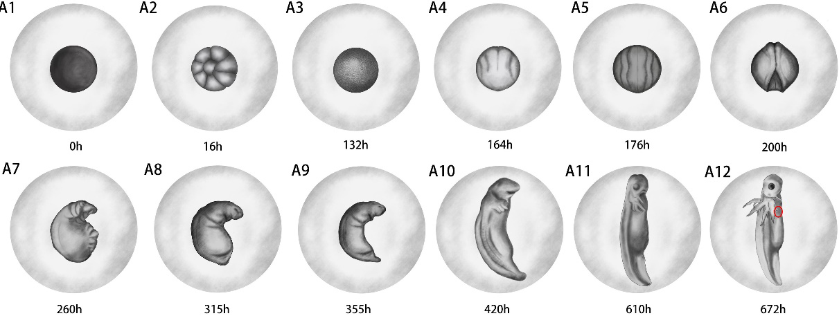
At 315 ± 3 h of embryonic development, the embryo elongated along the neural tube toward both ends, tail buds appeared, and both the primitive head and tail buds curved ventrally, with a more pronounced expansion of the abdomen, referred to as the early tail bud stage (Fig. 1 A8). The embryo’s body size increased, and there was a slight depression on both sides of the head. At 355 ± 5 h of embryonic development, the abdominal expansion became markedly smaller, and the tail bud contour was clear, referred to as the late tail bud stage (Fig. 1 A9). At 420 ± 5 h of embryonic development, the embryo was larger and more extended, the larval body was largely formed, and three primitive external gills and a balance bar appeared on both sides of the head, referred to as the early external gill stage (Fig. 1 A10).
At 610 ± 3 h of embryonic development, the embryonic head organ development was essentially complete. The eye was basically formed, and smaller gill filaments were present on the three pairs of gills, referred to as the late external gill stage (Fig. 1 A11). At 672 ± 3 h of embryonic development, as the embryo continued to develop, the hatchling’s morphology matured, the gill filaments on the three external gills became abundant, the balance rods were clearly visible, forelimb buds were forming, and the hatchling was occasionally seen to move within the egg, referred to as the incubation period (Fig. 1 A12). At 694 ± 48 h of embryonic development, larvae broke through the egg membrane and hatched out, usually with a small amount of yolk remaining. Most lay on their sides at the bottom of the water and are referred to as out-of-membrane larvae (Fig. 2 B1).
Anji salamander balance bar stage
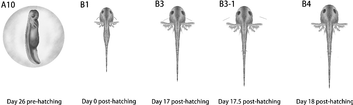
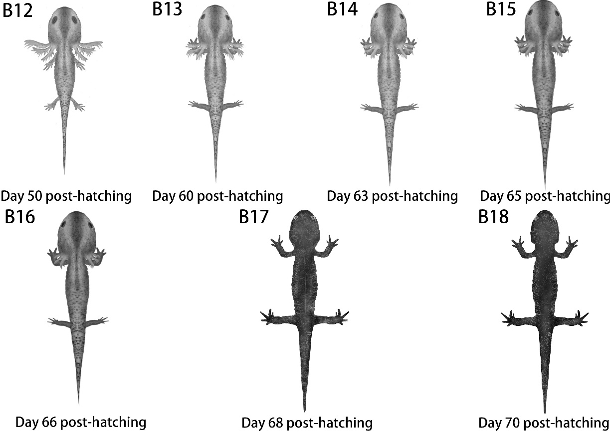
We define the day of emergence of hatch as post-hatching day 1. Balance bars began to appear and develop in the early stages of the external gills. Small bar-like tissues appeared on both sides, and the stage from the appearance of the balance bars to their complete shedding is referred to as the balance bar stage. The development of the balance bars on both sides of the head was completed in the embryo 2-3 days before exsanguination (Fig. 1 A10). After the membrane was removed, the balance bars gradually changed from a short, thick state to a long, slender form. This transformation essentially stopped around the 17.5 ± 2.5 day post-hatching, reaching a length of approximately 2.00 ± 0.50 mm, and disappeared by the 19 ± 2 day post-hatching (Fig. 3). The balance bars of Anji salamander disappeared in an asymmetric shedding pattern, and the balance bars were shed with either the left or right side of the bars shed first, and the other side shedding within the next 24 h. Moreover, the entire stage in which the balance bar exists is accompanied by the development of the forelimbs, and the hindlimbs began to develop later in the development of the balance bar.
On day 5 ± 2 post-hatching, the larvae began feeding, and if there was no external food as an opener, Anji salamander might resort to intraspecific predation. The swallowing process typically involved biting the head or other body parts, with the prey being gradually ingested. The digestive time varies depending on the size of the food, and some of the digestive time in the mouth could reach up to 16 hours or even longer. After hunting for a companion, Anji salamander hide in a quiet corner and slowly enjoyed a meal on its own. However, if it was attacked or otherwise disturbed, it would assess the risk of abandoning the food or taking it and running away. During this stage, Anji salamander was relatively active, preferring dark and quiet water or even hiding in the gaps between rocks or in the mud underwater, occasionally swimming out of the water. Individuals at this stage were small, flexible, and preferred to be alone. They exhibited agitation during balance bar shedding and gradually settled down afterwards.
Anji salamander limb developmental stage
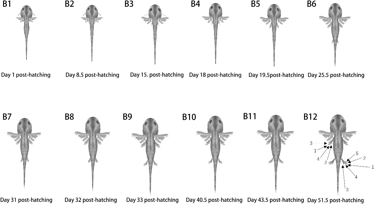
The process of limb development, including the 4 fingers on the forelimbs and the 5 toes on the hindlimbs, from their emergence to complete development, is referred to as the limb developmental stage of Anji salamander. As early as the embryonic stage, the forelimb bud began to develop (Fig. 1 A12). On day 8.5 ± 1.5 post-hatching, the forelimb bud developed into the forearm (Fig. 2 B2). On day 15.5 ± 1.5 post-hatching, the first forelimb finger began to differentiate, but the longest clear skin was followed by the development of the fourth finger (Fig. 2 B3). On day 18.5 ± 1.5 post-hatching, the second finger of the forelimb began to differentiate (Fig. 2 B4). On day 18 ± 2 post-hatching, the hindlimb bud also began to form (Fig. 2 B4). On day 19.5 ± 1.5 post-hatching, the forelimb’s third finger and the hindlimb’s forearm began to develop (Fig. 2 B5). On day 20.5 ± 1.5 post-hatching, the fourth finger of the forelimb began to differentiate, and capillaries started to appear within the longest transparent skin layer. On day 25.5 ± 1.5 post- hatching, the rudimentary form of the four fingers was formed, but there were still web-like connections between each finger (Fig. 2 B6).
On day 30 ± 1 post- hatching, the first toe of the hind limb began to differentiate (Fig. 2 B6). On day 31 ± 1 post- hatching, the second toe of the hind limb began to differentiate (Fig. 2 B7). On day 32 ± 1 post-hatching, the third toe of the hind limb began to differentiate, and at the same time, the skin of the fourth toe had also formed, but it had not yet undergone differentiation from capillaries (Fig. 2 B8). On day 33 ± 1 post-hatching, the fourth toe of the hind limb began to differentiate (Fig. 2 B9). On day 36.5 ± 1.5 post-hatching, the forelimbs had fully developed, with the fourth toe having formed and there were no web-like connection between the fourth toe and the other toes (Fig. 2 B9). On day 40.5 ± 1.5 post-hatching, the hind limb began to differentiate and the fifth toe emerged (Fig. 2 B10). On day 43.5 ± 1.5 post-hatching, the rudimentary form of the hind limb’s five toes was formed, although there were still web connections between the five toes of the hind limb (Fig. 2 B11). On day 51.5 ± 1.5 post-hatching, limb development was completed. At this point, the hind limbs had developed the fifth toe and there was no web connection between the five toes (Fig. 2 B12).
On day 34 ± 1 post-hatching, the ventral costal fovea begins to appear (Fig. 4 B9). On day 49 ± 1 post-hatching, the ventral costal fovea gradually became deeper (Fig. 4 B10). The four fingers of the forelimbs of Anji salamander developed in the following sequence from top to bottom: 3, 1, 4, 2 (Fig. 2 B12). That is, the finger at the top of the picture developed as the third one, followed by the first, fourth and second fingers in sequence. The sequential development order of the five toes of the hind limbs from top to bottom was 3, 4, 1, 2, 5 (Fig. 2 B12).
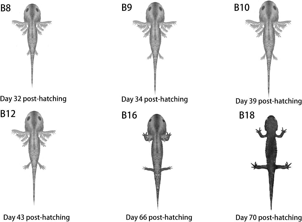
The behavioral characteristics during this stage were mainly characterized by using live prey as the main food source, with an increase in movement ability. They could swim, or move on the bottom of the water, or reach the surface. The growth rate was relatively fast. During this stage, if the larval Anji salamander lived in groups and touched each other’s bodies, they would engage in mutual attacks and biting, especially on the limbs and external gills. By this stage, the limbs, the four pairs of fingers on the front limbs and the five pairs of toes on the hind limbs had fully developed, with the body length reaching 50.0 ± 5.0 mm.
Anji salamander pre-metamorphosis stage
The stage from the completion of the development of the limbs and fingers of the Anji salamander to 12 ± 3 days before the metamorphosis stage is referred to as the pre-metamorphosis stage. During this stage, the gill filaments of Anji salamander turned red, the blood vessels became abundant, the body size increased larger and longer, the limbs became strong and powerful, the body became plump, with a body length of 55.0 ± 5.0 mm. The dorsal fin gradually disappeared, the ventral costal fovea deepened, and the eyeballs slightly protruded.
The behavioral characteristics during this stage are mainly characterized by a significant increase in food intake, frequent movement at the bottom of the water and in the upper water layer, and sometimes extending their heads close to the water surface to exhale bubbles. They prefer to stay in dark corners alone, had less activity during the day, and were relatively more active at night.
Anji salamander metamorphosis stage
The external gills began to retract inward until they completely disappeared. The breathing method changed from gill respiration to using skin and lungs for breathing, and the lifestyle shifted from aquatic to semi-aquatic. This process is what we call the metamorphosis period. During the metamorphic stage, the gill filaments of Anji salamander gradually became thicker and shorter, and the external gills also became shorter (Fig. 5), and before the external gills had completely disappeared, Anji salamander began to undergo its first molting process of life (Fig. 6). Subsequently, the skin on the upper part of the gill cover and the skin on the neck gradually merged and covered the area where the gills had been exposed, forming a neck fold. Eventually, the outer gills completely disappeared. During this stage, the head of Anji salamander became more pointed, its body color darkened, white spots appeared on its body surface. There was a significant protrusion of the eyeball when compared to the larval eyeball, and the ventral costal fovea continued to deepen.
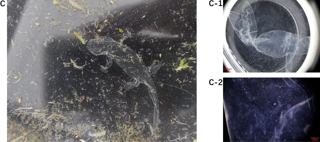
The behavioral characteristics of Anji salamander during its metamorphosis stage were that it spent a long time hiding in darker places, was sensitive to light and sound, had a darker body color and more body spots, with enhanced color-changing and concealment abilities. During metamorphosis stage, there was almost no phenomenon of mutual attacks or cannibalism. After the external gills disappeared, Anji salamander began to engage in long-distance crawling, leaving the water puddles and searching for new habitats and food. Its dietary characteristics also underwent significant changes. From the early stage of metamorphosis, its appetite significantly weakened and even led to prolonged fasting. It took approximately 42 ± 3 days of fasting before it could resume normal feeding.
Anji salamander subadult stage
We refer the period from the end of metamorphosis to sexual maturity as subadult stage. During this stage, the color of Anji salamander was significantly different from that of the larvae. The color of the subadult is darker and has white dot-like spots compared to the larvae. The head of the larva was sharper, the eyes were more prominent and had fully formed ventral costal fovea (Fig. 5 B12, B18). During the subadult stage, the body length and weight of the salamander gradually increased, and it underwent frequent molting. The aging skin usually fell off from the head to tail, forming a complete “shell” (Fig. 6C). Under the microscope, the shed skin maintained the body’s shape (Fig. 6 C, C-1), and the pigment spots on the skin might also be clearly observed (Fig. 6 C-2). The entire exfoliation process took approximately 12 hours.
In terms of behavior, Anji salamander at this stage seemed to prefer living in groups, especially shortly after their abnormal development. There was almost no phenomenon of cannibalism. Groups of three or four can be seen gathering together. However, they had territorial awareness. If their territory was invaded by outsiders, there might be biting behavior, mostly targeting the limbs and tail. During this stage, although they could live away from water, they still required a high level of humidity. If they left the water environment and the humidity was insufficient, they might suffer from dehydration, leading to skin wrinkling, tail curling, and in severe cases, it could even endanger their lives.
General characteristics of the development from fertilized egg to subadult stage of Anji salamander
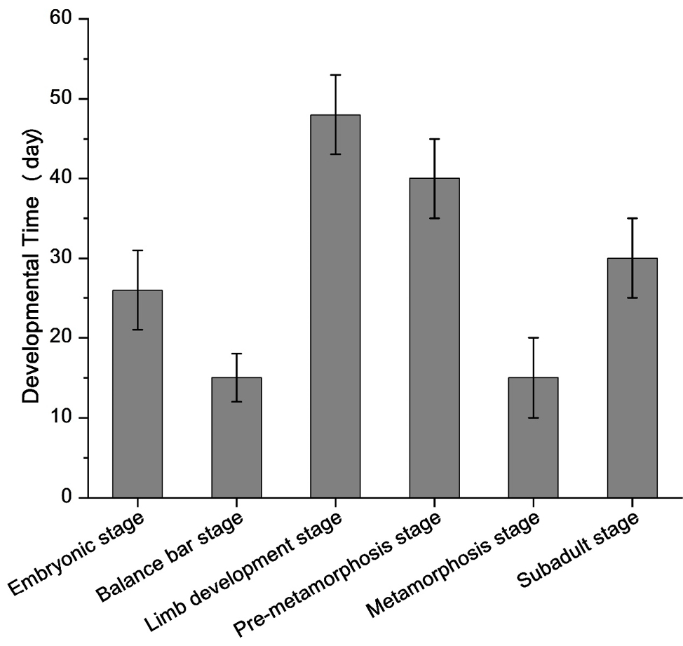
In aquatic environments at 4-15 °C, the embryonic development period of Anji salamander lasted for 30 ± 5 days. The higher the temperature, the faster the development. From the fertilized egg to the subadult stage, there were a total of six major developmental stages: embryonic development stage, balance bar stage, limb development stage, pre-metamorphosis stage, metamorphic stage, and subadult stage. The balance bar stage required 18 ± 3 days, the limb development stage 50 ± 5 days, the pre-metamorphosis stage 10 ± 5 days, the metamorphosis stage 10±5 days, and after the metamorphosis is completed, it took 30 ± 5 days to develop into subadult stage (Fig. 7).
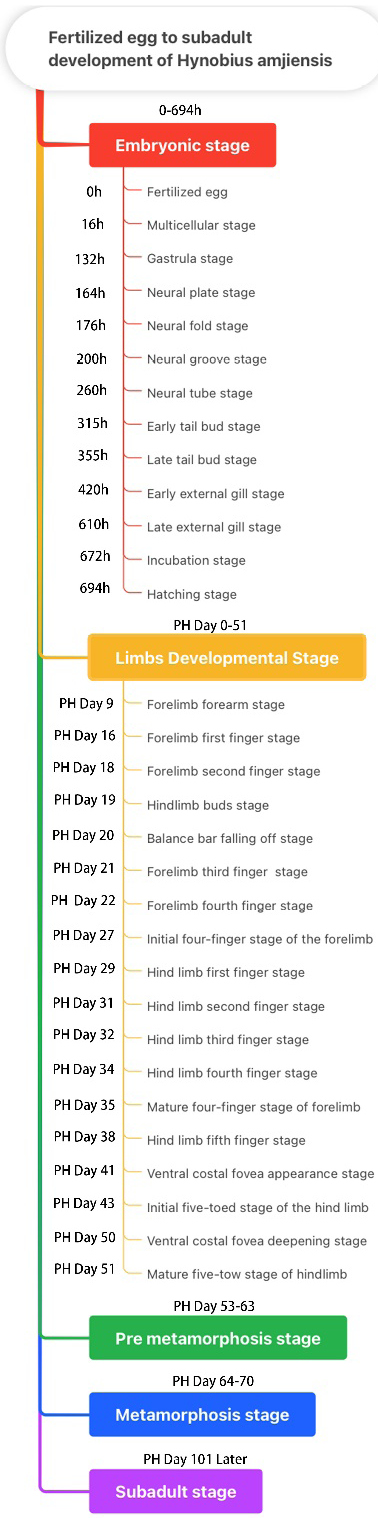
There were 34 developmental nodes, which are Fertilized egg, Multicellular stage, Gastrula stage, Neural plate stage, Neural fold stage, Neural groove stage, Neural tube stage, Early tail bud stage, Late tail bud stage, Early external gill stage, Late external gill stage, Incubation stage, Hatching stage, Forelimb forearm stage, Forelimb first finger stage, Forelimb second finger stage, Hindlimb buds stage, Balance bar falling off stage, Forelimb third finger stage, Forelimb fourth finger stage, Forelimb four - finger initial stage, Hind limb first finger stage, Hind limb second finger stage, Hind limb third finger stage, Hind limb fourth finger stage, Forelimb four-finger mature stage, Hind limb fifth finger stage, Ventral costal fovea appearance stage, Hindlimb five - toe initial stage, Ventral costal fovea deepening stage, Hindlimb five - toe mature stage, Pre metamorphosis stage, Metamorphosis stage, and Subadult stage (Fig. 8). The changes and characteristics of the 34 developmental nodes throughout the entire development process were presented in Table 1 and Table 2.
| Number | Developmental stage | Distinguishing Characteristics | Time of Emergence /h | Growth temperature/°C |
|---|---|---|---|---|
| 1 | Fertilized egg | The fertilized eggs are spherical, with a grayish-black color | 0 | 6.5 ± 3.5 |
| 2 | Multicellular stage | Cells continuously divide and the number of cells increases | 16 ± 3 | 6.5 ± 3.5 |
| 3 | Gastrula stage | The embryo is irregular and round, the embryo surface is smooth, and the blastoderm cells are involved into the embryo, with bulges or depressions on the edge | 132 ± 4 | 6.5 ± 3.5 |
| 4 | Neural plate stage | Embryo exhibits slight folding, shallow ditch appears | 164 ± 4 | 6.5 ± 3.5 |
| 5 | Neural fold stage | Embryonic folds became significantly opened | 176 ± 3 | 6.5 ± 3.5 |
| 6 | Neural groove stage | Embryonic folds were significantly elevated and converged to the center | 200 ± 3 | 6.5 ± 3.5 |
| 7 | Neural tube stage | Embryo is spindle shaped | 260 ± 5 | 6.5 ± 3.5 |
| 8 | Early tail bud stage | Embryonic head rudiments appear | 315 ± 3 | 6.5 ± 3.5 |
| 9 | Late tail bud stage | Embryonic eye sacs, gill arch, and tails are well characterized | 355 ± 5h | 6.5 ± 3.5 |
| 10 | Early external gill stage | Embryo appear with primitive outer gills and balance bars | 420 ± 5 | 6.5 ± 3.5 |
| 11 | Late external gill stage | Embryonic eye formation with three outer gills and gill filament development | 610 ± 3 | 6.5 ± 3.5 |
| 12 | Incubation stage | Embryonic development is basically complete, with a pair of balance rods and forelimb buds | 672 ± 3 | 6.5 ± 3.5 |
| 13 | Hatching stage | The larvae lie on their side under the water with a small amount of yolk residue | 694 ± 48 | 6.5 ± 3.5 |
| Number | Developmental stage | Distinguishing Characteristics | Time of Emergence /Day | Growth temperature/°C |
|---|---|---|---|---|
| 1 | Forelimb forearm stage | Larval forelimbs become longer and there is no differentiation of fingers | 8 ± 2 | 12 ± 3 |
| 2 | Forelimb first finger stage | Larval forelimbs fingers form a triangle | 13.5 ± 1.5 | 12 ± 3 |
| 3 | Forelimb second finger stage | Larval forelimbs fingers are bifurcated, emergence of hindlimb buds and the shedding of balance bars | 18.5 ± 1.5 | 12 ± 3 |
| 4 | Hindlimb buds stage | A pair of transparent and spotless buds appeared in the hindlimb | 18 ± 2 | 12 ± 3 |
| 5 | Balance bar falling off stage | The balance bar on both sides of the mouth became thinner and disappeared in the form of falling off | 19 ± 2 | 12 ± 3 |
| 6 | Forelimb third finger stage | Larval forelimb fingers are trifurcat | 19.5 ± 1.5 | 12 ± 3 |
| 7 | Forelimb fourth finger stage | Larval forelimb fingers are basically formed, there are webbed connections between the fingers | 20.5 ± 1.5 | 12 ± 3 |
| 8 | Forelimb four - finger initial stage | Larval forelimbs with four fingers basically formed and webbed connections between fingers | 21.5 ± 1.5 | 13 ± 3 |
| 9 | Hind limb first finger stage | Larva hind limb bud is triangular | 30 ± 1 | 13 ± 3 |
| 10 | Hind limb second finger stage | Larval hind limbs toes are quad,but only two toes developed, and the excess was cutis | 31 ± 1 | 13 ± 3 |
| 11 | Hind limb third finger stage | Larval hind limbs toes are quad, but only three toes developed, and the excess was cutis | 32 ± 1 | 13 ± 3 |
| 12 | Hind limb fourth finger stage | Larval hind limbs are four to four forked | 33 ± 1 | 13 ± 3 |
| 13 | Forelimb four-finger mature stage | Larval forelimb fingers are fully developed, and there is no web connection between the fingers | 36.5 ± 1.5 | 13 ± 3 |
| 14 | Hind limb fifth finger stage | Larval hind limbs have pentamerous toes | 40.5 ± 1.5 | 13 ± 3 |
| 15 | Ventral costal fovea appearance stage | Larval appear as indistinct depressions on both sides of the abdomen near the head | 42 ± 1 | 13 ± 3 |
| 16 | Hindlimb five - toe initial stage | Larval five toes of the hind limbs of juveniles are largely formed, but are connected by webbing between the toes | 43.5 ± 1.5 | 13 ± 3 |
| 17 | Ventral costal fovea deepening stage | Larvae have more defined depressions on both sides of the abdomen and develop towards the sides of the abdomen near the hind limbs | 44 ± 1 | 13 ± 3 |
| 18 | Hindlimb five - toe mature stage | Five toes of the hind limb form but there are webbed connections between the toes | 51.5 ± 1.5 | 13 ± 3 |
| 19 | Pre metamorphosis stage | The bulge of the larva’s eyeball, the body color becomes darker, and the caudal fin becomes smaller | 58 ± 5 | 14 ± 1 |
| 20 | Metamorphosis stage | The three outer gills of the larva gradually shorten, the body color deepens, and a small amount of white spots appear | 67 ± 4 | 14 ± 1 |
| 21 | Subadult stage | No outer gills, increased white spots, neck folds | 106 ± 5 | 14 ± 1 |
Discussion
Embryonic development of Anji salamander
The egg capsule of Anji salamander could develop normally under normal conditions without any infection, except for a few unfertilized eggs. The unfertilized eggs would gradually disintegrate, and the cells eventually died as the eggs became concave. The embryonic development process of Anji salamander is extremely similar to that of other Hynobiidae species, including other vertebrates. For instance, Hynobius leechii, Hynobius guabangshanensis, Hynobiid Salamander, Onychodactylus japonicus, and fish all undergo the developmental process of fertilized eggs, cleavage, blastula, gastrula, organogenesis, and finally membrane formation (Iwasawa and Kera, 1980; Iwasawa and Yamashita, 1991; Ma et al., 1994; Mi et al., 2008; Mousavi et al., 2022). The difference is that the forelimbs of Anji salamander were merely a raised bud-like structure before hatching, while those of the Chinese salamander have already developed into a bifurcated shape and have formed the rudimentary form of two fingers before hatching.
The development time varies even among different species and within the same species. During the embryonic development stage, the development time we measured is slightly different from two recent reports (Cao et al., 2024; Qiu et al., 2024). This might be due to environmental factors, such as light exposure, water temperature, oxygen saturation, etc., resulting in different developmental speeds. Under natural conditions, the development speed of the eggs laid by Anji salamander varied at different times. Generally, eggs incubated in warmer water developed more rapidly.
The balance bar of Anji salamander
In other species of Hynobius, the presence of balance bars has been observed during a similar stage (Zhang et al., 1987b; Ma et al., 1994; Mi et al., 2008). Furthermore, Ning et al. (2021) also discovered on Hynobius maoershanensis that the balance bar will fall off (Ning et al., 2021). In this study, we not only reported the times of the appearance and disappearance of the balance bar but also the fact that the balance bar fell out in an asymmetric manner. This asymmetric detachment might imply that the development of the balance bars on the left and right sides was not synchronous.
So far, there have been no relevant studies on the function of the balance bar in salamanders. We hypothesize that the balance bars of Anji salamander are similar to the lateral lines and antennae of other vertebrates, which serve the functions of navigation, detecting and sensing changes in the aquatic environment, intra-species communication to assist in locomotor equilibrium, and predation, etc. Lateral line exists in fish, primarily composed of the superficial and optic nerve mounds (Omura et al., 2014). The functions of the lateral line in fishes include sensing changes in the water flow and pressure (Mogdans, 2019), as well as prey detection, spatial localization, predator avoidance, flocking behavior, intraspecific communication, and station keeping (Klein and Bleckmann, 2015). Catfish have tentacles which are sensitive to changes in water flow and help catfish navigate, sense and hunt in dark environments (Biedenbach, 1971; Muka et al., 2010). Lateral line is also present in axolotls (Northcutt et al., 1994). We found Anji salamander larvae were able to sense food beyond the distance of the balance bar and slowly swim towards the prey, quickly biting it when the bar was about to touch or touching. At the same time, if another salamander leaned into another salamander, they were both able to quickly avoid each other without touching their bodies.
Patterns of limb development in the Anji salamander
There are two patterns of vertebrate limb development. One pattern is preaxial dominance, where structures close to the medial side of the body (i.e., radial or thumb side) preferentially form or differentiate during the development of an animal’s limbs, such as the radius develops before the ulna in forelimb development, and the 2nd finger develops before the 4th finger (Royle and Young, 2021). The other is postaxial dominance, where structures close to the lateral side of the body (i.e., ulnar or pinky side) preferentially form during the development of an animal’s limbs (Shubin, 1986). The limb development of Anji salamander follows a preaxial dominance developmental pattern, similar to that of many other Caudata (Fröbisch et al., 2007). In terms of the morphology of the limbs, except for the Salamandrella keyserlingii (Zhang et al., 1993c) and Batrachuperus pinchonii (Jia et al., 2018) which have 4 fingers on the forelimbs and 4 toes on the hindlimbs, most species of Hynobiidae have 4 fingers on the forelimbs and 5 toes on the hindlimbs (Ma et al., 1994; Mi et al., 2007; Ning et al., 2021). The preaxial dominance developmental pattern may reflect its long-term environmental adaptation. It may also be closely related to the evolutionary process of 4-toed to 5-toed hindlimbs in the Hynobiidae (Ledbetter et al., 2019; Jia et al., 2022; Urošević et al., 2024). It has been shown that the four fingers of the forelimbs of amphibians with Caudata are adapted to aquatic life, which helps to reduce the resistance when swimming, and the forelimbs also have the role of maintaining the balance and stability of the body, while the five toes of the hindlimbs are adapted to terrestrial life and used to support the body and propulsion. These forelimbs and hindlimbs characteristics are highly compatible with their amphibious lifestyle (Urošević et al., 2024).
We observed that the front four limbs of Anji salamander gathered to form a shovel-like grubbing away soil or peat moss, similar to digging a burrow, a process in which the back five fingers grasped the ground to support the body. We hypothesize that the 4-fingered front and back salamanders and the 4-fingered front and 5-fingered back salamanders evolved different numbers of fingers due to differences in their lifestyles, such as some salamanders need to dig burrows, while others do not; some live in static waters, while others inhabit dynamic waters. The corresponding number of fingers allows them to better adapt to the resistance of water currents in their environments. Further data are needed to validate or refute this hypothesis.
The limb function of Hynobius amjiensis may serve the same purposes of assisted locomotion and maintenance of balance as many vertebrates (Molnar et al., 2021; Smith et al., 2021; Chen et al., 2025b). In the larval stage, when there are individual small salamanders with bitten limbs, their balance and locomotor ability will be reduced, and their swimming speed will be slowed down or even flipped on its side, so the limbs in the larval stage may play an important role in their locomotion and balance. The development of forelimb buds in Hynobius amjiensis larvae prior to membrane the emergence (Fig. 1 A12) may be an attempt to adapt as quickly as possible to body the balance in the water after emergence from the membrane. Following the disappearance of the balance bars, both hind limbs developed rapidly (Fig. 3 B4), which allowed the body to continue to have “four symmetrical supports”, which may have been important for the maintenance of the body’s locomotor equilibrium. After the development of the four limbs is completed, Hynobius amjiensis larvae often crawl underwater, probably to develop the support and propulsion function of the hind limbs, which lays the foundation for the metamorphosis to crawl long distances to the shore in search of food and habitats. It has been suggested that the forelimbs of tetrapods mainly play a braking role and the hindlimbs mainly play a propulsive role (Kawano and Blob, 2013). Furthermore, we observed in the field that Hynobius amjiensis may have crawled out of underwater burrows to pair-lay their eggs (Appendix 1). We hypothesized that their more pointed heads and stout limbs may have the ability to excavate the burrows, but we need further studies to confirm our speculations.
Molting of Anji salamander
This is the first report of the molting phenomenon in Anji salamander. Molting is a natural process of growth and development in vertebrates, influenced by environmental and hormonal factors (Jorgensen et al., 1960; Heimeier and Shi, 2010). Extensive studies have shown that the molting process of amphibians is regulated by thyroid hormones (Dowling et al., 1966; Tata, 2006). It has been found that the level of thyroid hormone secretion in amphibians increases during metamorphosis (Denver, 2021; Tanizaki et al., 2021). The first molt occurred during the metamorphosis stage, and it is possible that hormones were also involved in the development of molt and metamorphosis in Anji salamander.
Amphibian molting behavior is necessary for their metamorphosis. The main component of molting is the cuticle of the skin surface, and the presence of molting indicates some keratinization of the skin (Heimeier and Shi, 2010; Akat et al., 2023). The skin of amphibians serves as an important organ for their respiration and gas exchange (Tattersall, 2007), and molting might regulate respiration and reduce water loss to some extent (Torri et al., 2020). It has also been implied that amphibians in the growth phase molt as a result of their body growth becoming increasingly tighter with their cuticle, and the old cuticle skin needs to be shed to accommodate growth (Lu, 2018). Molting can also defend against pathogenic microorganisms on the old skin, removing pathogenic microorganisms that have accumulated on the old skin over time (Ohmer et al., 2017). In this study, we found that Anji salamander molted more frequently after completing metamorphosis, so the molt of Anji salamander might be more closely related to their growth, development, immunity and other physiological processes.
Risks faced during metamorphosis and development of Anji salamander
Amphibian metamorphosis enables amphibians to cross the formidable ecological barrier between aquatic and terrestrial environments by coordinating morphological remodeling with physiological functional transitions, a process critically important for their subsequent survival. Studies have indicated that during amphibian metamorphosis, the degeneration of external gills may exhibit temporal mismatch with pulmonary developmental processes. Furthermore, the gradual functional decline in gill respiratory capacity throughout metamorphic progression can potentially lead to hypoxic stress and associated mortality during gill resorption (Burggren, 1982; 1994). Amphibians use their skin to assist respiration before and after gill shedding. However, after metamorphosis, the use of skin for respiration needs to be carried out at a more suitable humidity; otherwise, dry environments may cause respiratory distress, dehydration, and disruption of skin microflora, increasing the risk of infection with pathogenic microorganisms (Akat et al., 2023; Buttimer et al., 2024; Riddell et al., 2024). Amphibians can experience long period of cessation of feeding during metamorphosis due to multiple reasons, including hypothalamic regulation and reorganization of the digestive system (Kuzmin, 1997; Bender et al., 2018; Zhu et al., 2020).
Anji salamanders spend the vast majority of their metamorphosis to subadult in a state of relative feeding cessation, and have to face threats such as hypoxia from respiratory shifts after emerging onto land, significant energy expenditure from crawling long distances, and elevated habitat temperatures, and even failing to survive once they mistakenly enter a dry area with water-starved and wrinkled skin and frizzled tails for 12-24 hours. Studies have also been shown that some small salamanders maintain a larval form and are unable to complete metamorphosis (Okamiya et al., 2021). Additionally, skin fusion of the head and neck following gill internalization might greatly burden the immune and endocrine systems. Pathogen invasion at this stage could pose a significant survival threat. Therefore, we believe that the metamorphosis stage is a high-risk stage from fertilized eggs to subadults in Anji salamander.
Conclusions
In this study, we delineated the six main developmental processes of Anji salamander (Fig. 8) encompassing the whole developmental process, including 34 developmental nodes (Tables 1 and 2), and described the life habits of different developmental periods, filling gaps in the life history of Anji salamander. It is worth noting that Anji salamander is particularly vulnerable during the balance bar shedding stage and throughout metamorphosis, which is informative for conservation. For example, we in the reserve may consider the need to keep the habitat as free from external disturbance as possible before and after the shedding of the balance bar and during the process of metamorphosis. In addition, the subadult stage of Anji salamander has a relatively high dependence on air humidity and water. Therefore, if there is an extreme drought before or after the Anji salamander comes ashore, we can take the necessary intervention to ensure adequate water supply in the reserve. In conclusion, the knowledge of the life history of Anji salamander from larvae to subadults is useful for its conservation and future captive breeding.
Acknowledgments
We would like to express our gratitude to Anji Huangpu Jiangyuan Tourism Development Co., Ltd. for their financial support to this public welfare research. We thank the Animal Welfare Ethical Committee and Animal Experimental Ethics Committee of the China Huzhou University Research Institute for approving our work.
References
Akat Çömden, E., Yenmiş, M., Çakır, B. (2023): The complex bridge between aquatic and terrestrial life: Skin changes during development of amphibians. J. Dev. Biol. 1: 6.
Biedenbach, M.A. (1971): Functional properties of barbel mechanoreceptors in catfish. Brain Res. 27: 360-364.
Burggren, W.W., West, N.H. (1982): Changing respiratory importance of gills, lungs and skin during metamorphosis in the bullfrog Rana catesbeiana. Respir. Physiol. 47: 151-164.
Begon, M., Harper, J.L., Townsend, C.R. (1986): Ecology: Individuals, populations and communities, pp. 130-330. Wiley-Blackwell, Hoboken, New Jersey, USA.
Burggren, W.W., Infantino, R.L. (1994): The respiratory transition from water to air breathing during amphibian metamorphosis. Integr. Comp. Biol. 34: 238-246.
Bender, M.C., Hu, C., Pelletier, C., Denver, R.J. (2018): To eat or not to eat: Ontogeny of hypothalamic feeding controls and a role for leptin in modulating life-history transition in amphibian tadpoles. Proc. R. Soc. B Biol. Sci. 285: 20172784.
Buttimer, S., Moura-Campos, D., Greenspan, S.E., Neely, W.J., Ferrante, L., Toledo, L.F., Becker, C.G. (2024): Skin microbiome disturbance linked to drought-associated amphibian disease. Ecol. Lett. 27: e14372.
Chen, C.S., Yang, J., Yu, L.P., Guo, R. (2018): Reproductive biology of the critically endangered Anji salamander (Hynobius amjiensis), 59 pp. Proceedings of the Thirteenth Membership Congress and Academic Symposium of the Zoological Society of Zhejiang Province, China, November 2018. Zhejiang Nature Museum, Longwangshan Nature Reserve Administration, Qingliangfeng National Nature Reserve Administration, Eds., China.
Cao, Z., Guo, R., Fang, Z., Wang, Z., Liu, Y., Lin, L., Ji, X. (2024): Normal table of embryonic development in the Anji salamander Hynobius amjiensis (Hynobiidae). Dev. Biol. 511: 84-91.
Chen, C.S., Jia, J., Wang, X.T., Yang, J., Gao, K.Q. (2025): Anatomy of the critically-endangered Anji salamander (Hynobius amjiensis) provides new insights into morphological evolution of salamanders. J. Morphol. 286: e70028.
Dowling, J.T., Razevska, D. (1966): Thyroxine metabolism by amphibian skin during metamorphosis and molting. Gen. Comp. Endocrinol. 6: 162-169.
Denver, R.J. (2021): Stress hormones mediate developmental plasticity in vertebrates with complex life cycles. Neurobiol. Stress. 14: 100301.
Fröbisch, N.B., Carroll, R.L., Schoch, R.R. (2007): Limb ossification in the Paleozoic branchiosaurid Apateon (Temnospondyli) and the early evolution of preaxial dominance in tetrapod limb development. Evol. Dev. 9: 69-75.
Fröbisch, N.B., Bickelmann, C., Olori, J.C., Witzmann, F. (2015): Deep-time evolution of regeneration and preaxial polarity in tetrapod limb development. Nature. 527: 231-234.
Gu, H.Q., Ma, X.M., Wang, J., Du, Z.H., Lou, X.Q. (1999): Population size and population dynamics of the small Anji salamander (Hynobius amjiensis). Sichuan Zool. 18:104-106.
Heimeier, R.A., Shi, Y.B. (2010): Amphibian metamorphosis as a model for studying endocrine disruption on vertebrate development: Effect of bisphenol A on thyroid hormone action. Gen. Comp. Endocrinol. 168: 181-189.
Iwasawa, H., Kera, Y. (1980): Normal stages of development of the Japanese lungless salamander, Onychodactylus japonicus (Houttuyn). Jpn. J. Herpetol. 8: 73-89.
Iwasawa, H., Yamashita, K. (1991): Normal stages of development of a hynobiid salamander, Hynobius nigrescens Stejneger. Jpn. J. Herpetol. 14: 39-62.
Jorgensen, C., Larsen, L. (1960): Hormonal control of moulting in amphibians. Nature 185: 244-245.
Jia, J., Zhang, M.H., Gao, K.Q., Jiang, J.P. (2018): Dataset of μCT scan of the holotype specimen of Batrachuperus londongensis Liu and Tian, 1978, an endemic hynobiid (Amphibia, Urodela) from Mount Emei, Sichuan Province, China. Chin. Sci. Data. 3: 79-93.
Jia, J., Anderson, J.S., Jiang, J.P., Wu, W., Shubin, N.H., Gao, K.Q. (2022): Ossification patterns of the carpus and tarsus in salamanders and impacts of preaxial dominance on the fin-to-limb transition. Sci. Adv. 8: eabq7669.
Kuzmin, S.L. (1997): Feeding of amphibians during metamorphosis. Amphib.-Reptil. 18: 121–131.
Kawano, S.M., Blob, R.W. (2013): Propulsive forces of mudskipper fins and salamander limbs during terrestrial locomotion: Implications for the invasion of land. Integr. Comp. Biol. 53: 283-294.
Klein, A., Bleckmann, H. (2015): Function of lateral line canal morphology. Integr. Zool. 10: 111-121.
Liu, B.Q. (2016): A new distribution site of the rare and endangered amphibian, Hynobius amjiensis, discovered in our province. Zhejiang For. Sci. Tech. 36: 28-28.
Lu, X.B., Jiang, Q., Min, Y., Jiang, X., Yang, Z.P., Sun, L. S., Zhang, X. J. (2018): Delineation of the molt cycle and the effect of molt frequency on growth of Rhodophorus rosenbergii. Freshw. Fish. 48: 88-93.
Ledbetter, N.M., Bonett, R.M. (2019): Terrestriality constrains salamander limb diversification: Implications for the evolution of pentadactyly. J. Evol. Biol. 32: 642-652.
Luedtke, J.A., Chanson, J., Neam, K., Hobin, L., Maciel, A.O., Catenazzi, A., Borzée, A., Hamidy, A., Aowphol, A., Jean, A., Sosa-Bartuano, Á., Fong, G.A., de Silva, A., Fouquet, A., Angulo, A., Kidov, A.A., Muñoz Saravia, A., Diesmos, A.C., Tominaga, A., Shrestha, B., Stuart, S.N. (2023): Ongoing declines for the world’s amphibians in the face of emerging threats. Nature. 622: 308-314.
Ma, L.D., Ma, D.K., Gao, A. (1994): Early development of the small salamander (Hynobius amjiensis) from Northeastern China. J. Zool. 232: 46-51.
Mi, X.Q., Deng, X.J., Guo, K.Q., Niu, Y.D., Zhou, Y. (2007): Early embryonic development of Hynobius guabangshanensis. Sichuan Zool. 26: 377-378+239.
Mukai, Y., Tuzan, A.D., Lim, L.S., Yahaya, S.H. (2010): Feeding behavior under dark conditions in larvae of sutchi catfish Pangasianodon hypophthalmus. Fish. Sci. 76: 457-461
Mogdans, J. (2019): Sensory ecology of the fish lateral-line system: Morphological and physiological adaptations for the perception of hydrodynamic stimuli. J. Fish Biol. 95: 53-72.
Molnar, J.L., Hutchinson, J.R., Diogo, R., Clack, J.A., Pierce, S.E. (2021): Evolution of forelimb musculoskeletal function across the fish-to-tetrapod transition. Sci. Adv. 7: eabd7457.
Mousavi, S.E., Patil, J.G. (2022): Stages of embryonic development in the live-bearing fish, Gambusia holbrooki. Dev. Dyn. 251: 287-320.
Meletiadis, A., Di Nicola, M.R., Bovero, S., Favelli, M., Pezzolato, M., Grella, S., Rezza, G., Acutis, P.L. (2025): Prevalence of Batrachochytrium dendrobatidis in amphibians in northwestern Italy’s protected areas. Animals. 15: 157.
Northcutt, R.G., Catania, K.C., Criley, B.B. (1994): Development of lateral line organs in the axolotl. J. Comp. Neurol. 340: 480-514.
Ning, M.H., Chi, H., Chen, Y., Yang, B., Wu, Z.J., Huang, H.Y. (2011): A preliminary study on the ontogenetic characteristics of captive-bred Hynobius maoershanensis. Sichuan Zool. 40: 196-202.
Nettle, D., Frankenhuis, W.E. (2019): The evolution of life-history theory: A bibliometric analysis of an interdisciplinary research area. Proc. R. Soc. B Biol. Sci. 286: 20190040.
Omura, A., Anzai, W., Endo, H. (2014): Functional and morphological variety in trunk muscles of Urodela. J. Vet. Med. Sci. 76: 159-167.
Ohmer, M.E.B., Cramp, R.L., Russo, C.J.M., White, C.R., Franklin, C.E. (2017): Skin sloughing in susceptible and resistant amphibians regulates infection with a fungal pathogen. Sci. Rep. 7: 3529.
Okamiya, H., Sugime, R., Furusawa, C., Inoue, Y., Kishida, O. (2021): Paedomorphosis in the Ezo salamander (Hynobius retardatus) rediscovered after almost 90 years. Zool. Lett. 7: 14.
Pan, D.S., Pang, Y.W., Zhou, Y.Y., Huang, Y.X., Xu, J.F. (2020): Effects of climate changes on the habitat of Hynobius amjiensis based on meteorological and remote sensing data. J. Hangzhou Norm. Univ. Nat. Sci. Ed. 19: 323-328.
Pottier, P., Kearney, M.R., Wu, N.C., Gunderson, A.R., Rej, J.E., Rivera-Villanueva, A.N., Pollo, P., Burke, S., Drobniak, S.M., Nakagawa, S. (2025): Vulnerability of amphibians to global warming. Nature. 639: 954-961.
Qiu, Y., Chen, K., Mei, Y., Yang, J., Chen, C. (2024): Pre-embryonic period observation shows a unique reproductive strategy of the critically endangered Anji salamander (Hynobius amjiensis). Animals. 14: 3007.
Royle, S.R., Young, J.J. (2021): Developmental biology: A 5’Hoxd-Gli3 balance in tetrapod axial polarity. Curr. Biol. 31: R1487-R1490.
Riddell, E.A., Burger, I.J., Muñoz, M.M., Weaver, S.J., Womack, M.C. (2024): Amphibians exhibit extremely high hydric costs of respiration. Integr. Comp. Biol. 64: 366-376.
Stearns, S.C. (1977): The Evolution of Life History Traits: A critique of the theory and a review of the data. Annu. Rev. Ecol. Syst. 8: 145-171.
Shubin, N.H., Alberch, P. (1986): A Morphogenetic Approach to the origin and basic organization of the tetrapod limb. In: Evolutionary biology, pp. 319-387. Hecht, M.K., Wallace, B., Prance, G.T., Eds., Springer, Boston, MA.
Smith, S.K., Hilliard Young, V.K. (2021): Balancing on a limb: Effects of gravidity on locomotion in arboreal, limbed vertebrates. Integr. Comp. Biol. 61: 573-578.
Tata, J.R. (2006): Amphibian metamorphosis as a model for the developmental actions of thyroid hormone. Mol. Cell. Endocrinol. 246: 10-20.
Tattersall, G.J. (2007): Skin breathing in amphibians. In: Endothelial biomedicine, pp. 85-91. Aird, W.C., Ed, Cambridge: Cambridge University Press, United Kingdom.
Torri, C., Falini, G., Montroni, D., Fermani, S., Teta, R., Mangoni, A., Alibardi, L. (2020): Cholesterol derivatives make large part of the lipids from epidermal molts of the desert-adapted Gila monster lizard (Heloderma suspectum). Sci. Rep. 10: 17197.
Tanizaki, Y., Shibata, Y., Zhang, H., Shi, Y.B. (2021): Analysis of thyroid hormone receptor α-Knockout tadpoles reveals that the activation of cell cycle program is involved in thyroid hormone-induced larval epithelial cell death and adult intestinal stem cell development during Xenopus tropicalis metamorphosis. Thyroid. 31: 128-142.
Tan, W.C., Herrel, A., Rödder, D. (2023): A global analysis of habitat fragmentation research in reptiles and amphibians: What have we done so far?. Biodivers. Conserv. 32: 439-468.
Urošević, A., Budečević, S., Ljubisavljević, K., Tomašević Kolarov, N., Ajduković, M. (2024): Morphological variation, modularity and integration in the scapula and humerus of Lissotriton newts. J. Anat. 245: 97-108.
Williams, G.C. (1966): Natural selection, the costs of reproduction, and a refinement of Lack’s principle. Am. Nat. 100: 687-690.
Yang, J., Chen, C.S., Chen, S.H., Ding, P., Fan, Z.Y., Lu, Y.W., Yu, L.P., Lin, H. (2016): Population genetic structure of critically endangered salamander in China: Recommendations for conservation. Genet. Mol. Res. 15.
Zhang, L.S., Tang, G.Q. (1987): Some biological data on the Chinese small salamander (Hynobius chinensis). Chin. J. Zool. 22: 23-25.
Zhang, K.Q., Zhang, Z.H., Chen, Y.M. (1993): Research on the reproductive habits of the northern salamander. Chin. J. Zool. 28: 9-12.
Zhang, K., Jiang, W.D. (2010): Demystifying the “Anji salamander”. Zhejiang For. Sci. Technol. 30: 16-17.
Zhu, W., Chang, L., Zhao, T., Wang, B., Jiang, J. (2020): Remarkable metabolic reorganization and altered metabolic requirements in frog metamorphic climax. Front. Zool. 17: 30.